Giant cell arteritis (temporal arteritis)
Exam relevance: in 1 of more than 50 board exam reports · rank 93
- Synonyms
- Arteriitis temporalis, Morbus Horton, Arteriitis cranialis, Horton-Syndrom, giant cell arteritis, temporal arteritis, Horton disease
- Specialty
- Dermatology · Vascular skin disorders
- Images
- Histology 2 · Clinical 2
- In the app
- 1 flashcard
- Last updated
- 10/2026 · Dr. Pascal Bafteh
Contents
Images (4)
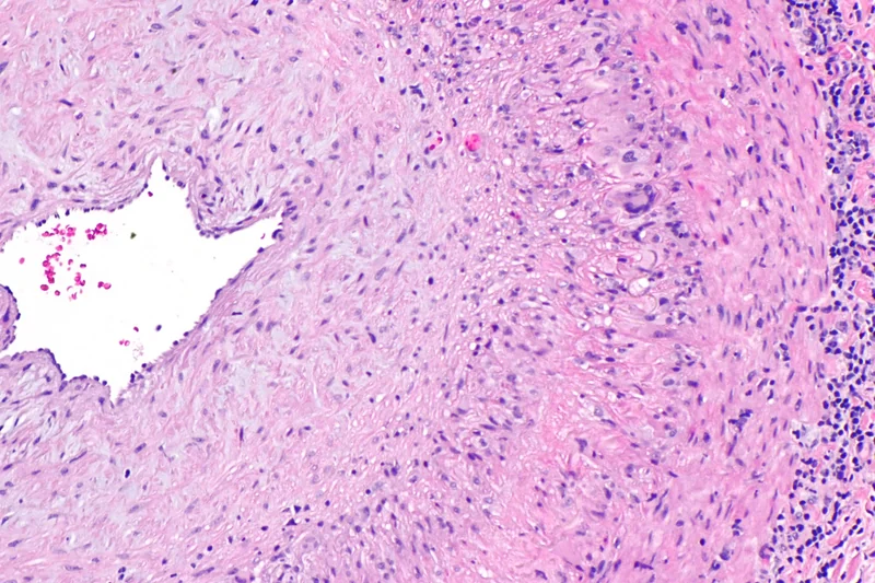 Histology
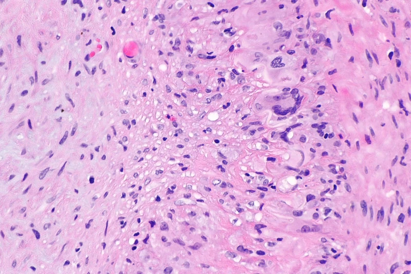 Histology
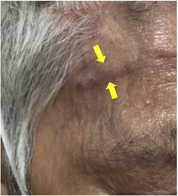
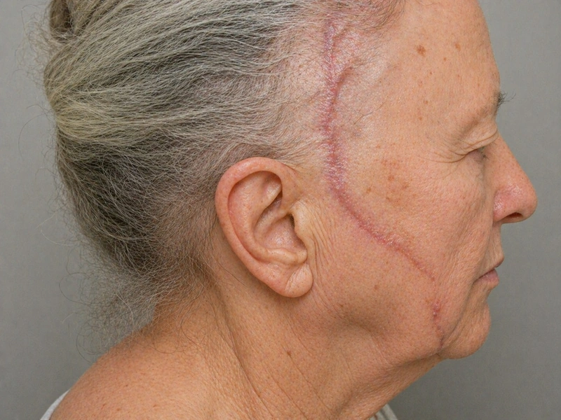 AI illustration
Definition
Giant cell arteritis is a granulomatous vasculitis of large and medium-sized arteries affecting the aorta and its branches, typically the extracranial branches of the carotid artery such as the superficial temporal artery. In Europe and the USA it is a relatively common form of vasculitis; intracranial vessels are usually not affected.
Occurrence & epidemiology
It occurs only in adults over 50, peaking between 70 and 80 years; women are affected more often than men, and people of European descent more often than others. In half of the cases polymyalgia rheumatica is present at the same time.
Clinical features
- New, persisting, often severe, sometimes throbbing headache (temporal, occipital, frontal or diffuse), tenderness and sensitivity of the scalp (e.g. when combing hair).
- Swollen, tender, sometimes nodular or erythematous temporal artery, sometimes with a diminished or absent pulse.
- Jaw claudication, constitutional symptoms such as fever, weight loss and fatigue.
- Visual disturbances (amaurosis fugax, diplopia, sudden visual loss due to anterior ischaemic optic neuropathy) – the most feared complication.
- Skin: rarely ischaemic necrosis and ulcers of the scalp in the territory of the temporal artery, also tongue necrosis.
Histology
Granulomatous inflammation of the arterial wall with mononuclear infiltrates (T cells, macrophages); multinucleated giant cells, when present, lie near the disrupted elastic lamina; marked intimal thickening with luminal narrowing. Inflamed and normal segments alternate ("skip lesions") – a negative biopsy therefore does not exclude the disease.
Diagnosis
- Inflammatory markers: ESR and CRP usually markedly raised.
- Colour duplex ultrasound of the temporal arteries (wall oedema seen as a "halo"), ideally performed early; performed by experts, it can replace biopsy in selected cases.
- Temporal artery biopsy for histological confirmation; additional imaging of the large vessels.
- American College of Rheumatology criteria, including age 50 or older, new localised headache, tenderness of the temporal artery and ESR above 50 mm/h.
Differential diagnoses
Treatment principles
- When the diagnosis is suspected, systemic glucocorticoids are started immediately because of the risk of blindness, without waiting for biopsy or imaging results.
- Biologics blocking the IL-6 receptor, now also JAK inhibitors, or conventional immunosuppressants are used to prevent relapse and spare steroids.
- Care is multidisciplinary with rheumatology and ophthalmology, including prophylaxis of steroid-related adverse effects.
Guideline: AWMF guideline register (no. 060-007)
More on this topic in the DermaFuchs app.
Keep learning in the app
References (selection)
Cross-references
More vascular skin disorders
- Great saphenous vein varicosity
- Stasis dermatitis (venous eczema)
- Deep vein thrombosis
- Leg ulcer (ulcus cruris)
- Urticarial vasculitis (normocomplementaemic and hypocomplementaemic)
- Antiphospholipid syndrome – skin manifestations (livedo racemosa, Sneddon syndrome)
- Cholesterol embolism (atheroembolism)
- Diabetic foot ulcer (malum perforans)
Keep learning: test yourself
One question from the DermaFuchs app on Giant cell arteritis (temporal arteritis) – tap an answer.
Question 1 of 1
What biopsy finding is characteristic of giant cell arteritis?
Answer
Correct: Granulomatous arteritis of the temporal artery. Granulomatous arteritis of the temporal artery is a characteristic finding in giant cell arteritis.
More questions like these – with explanations and spaced repetition – in the app.
Note: This page is intended for medical education and does not replace diagnosis or treatment decisions in individual cases.