Sjögren's syndrome
- Synonyms
- Sjogren syndrome, sicca syndrome, dry eyes and dry mouth, Sjögren disease, anti-Ro
- Specialty
- Internal medicine · Rheumatology & immunology
- Images
- Blood smear & cytology 1 · Clinical 1
- Last updated
- 10/2026 · Dr. Pascal Bafteh
Contents
Images (2)
 Blood smear & cytology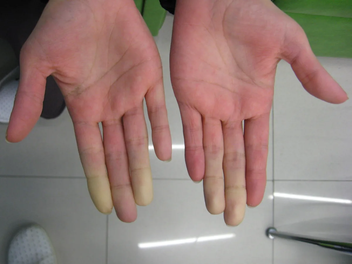
Definition
Sjögren syndrome is a chronic, systemic, autoimmune inflammatory disorder of unknown cause. Its hallmark is dryness of the mouth, eyes, and other mucous membranes (sicca syndrome) due to lymphocytic infiltration and dysfunction of exocrine glands. Numerous other organs may also be affected.
Classification
- Primary Sjögren syndrome: without another underlying disease.
- Secondary Sjögren syndrome: in about 30 % of people with autoimmune disorders such as rheumatoid arthritis, systemic lupus erythematosus, systemic sclerosis, mixed connective tissue disease, or Hashimoto thyroiditis.
2016 ACR/EULAR classification criteria (with at least one symptom of eye or oral dryness and no exclusion criteria; classification at 4 points):
- Focal lymphocytic sialadenitis in a labial salivary gland with focus score ≥ 1: 3 points
- Anti-SSA (Ro) antibodies: 3 points
- Ocular staining score ≥ 5 (or van Bijsterveld score ≥ 4) in at least one eye: 1 point
- Schirmer test ≤ 5 mm in 5 minutes in at least one eye: 1 point
- Unstimulated whole saliva flow rate ≤ 0.1 mL/min: 1 point
Occurrence & epidemiology
Middle-aged women are most frequently affected. Sicca symptoms without autoantibodies are more common than autoantibody-positive Sjögren syndrome.
Aetiopathogenesis
Genetic associations are known (e.g., HLA-DR3 in White people with primary Sjögren syndrome). Salivary, lacrimal, and other exocrine glands are infiltrated by CD4-positive T cells and B cells; cytokines such as interleukin-2 and interferon-gamma damage the ducts. Atrophy of the secretory epithelium of the lacrimal glands causes keratoconjunctivitis sicca; myoepithelial islands form in the parotid gland.
Clinical features
- Eyes: sandy or gritty sensation without itching; in advanced cases corneal damage (keratitis filiformis) with impaired vision.
- Mouth: xerostomia with difficulty chewing and swallowing, Candida infections, dental caries, salivary duct stones, diminished taste and smell.
- Dryness also of the skin, nose, throat, larynx, bronchi, and vagina; cough and dysphonia.
- Parotid enlargement in about one-third, usually firm, smooth, and mildly tender.
- Joints: arthralgias in about 50 %, nonerosive arthritis in about 20 %.
- Extraglandular manifestations: lymphadenopathy, Raynaud syndrome, interstitial lung disease, small-vessel vasculitis with purpura, polyneuropathy or mononeuritis multiplex, glomerulonephritis, distal renal tubular acidosis with hypokalemia, pancreatitis, fatigue.
- Lymphoma risk: about 16-fold increased risk of non-Hodgkin lymphoma, especially MALT lymphoma; indicators are low C4, monoclonal gammopathy, cryoglobulinemic vasculitis, and persistent parotid enlargement.
Diagnosis
- Eyes: Schirmer test (healthy about 15 mm wetting in 5 minutes, in Sjögren usually under 5 mm; about 15 % false-positive and 15 % false-negative results each), staining with rose bengal or lissamine green (ocular staining score), tear breakup time under 10 seconds on slit-lamp examination.
- Salivary glands: salivary flow measurement (≤ 0.1 mL/min), less often sialography or scintigraphy; simple clues are absent pooling of saliva under the tongue or a tongue blade sticking to the buccal mucosa.
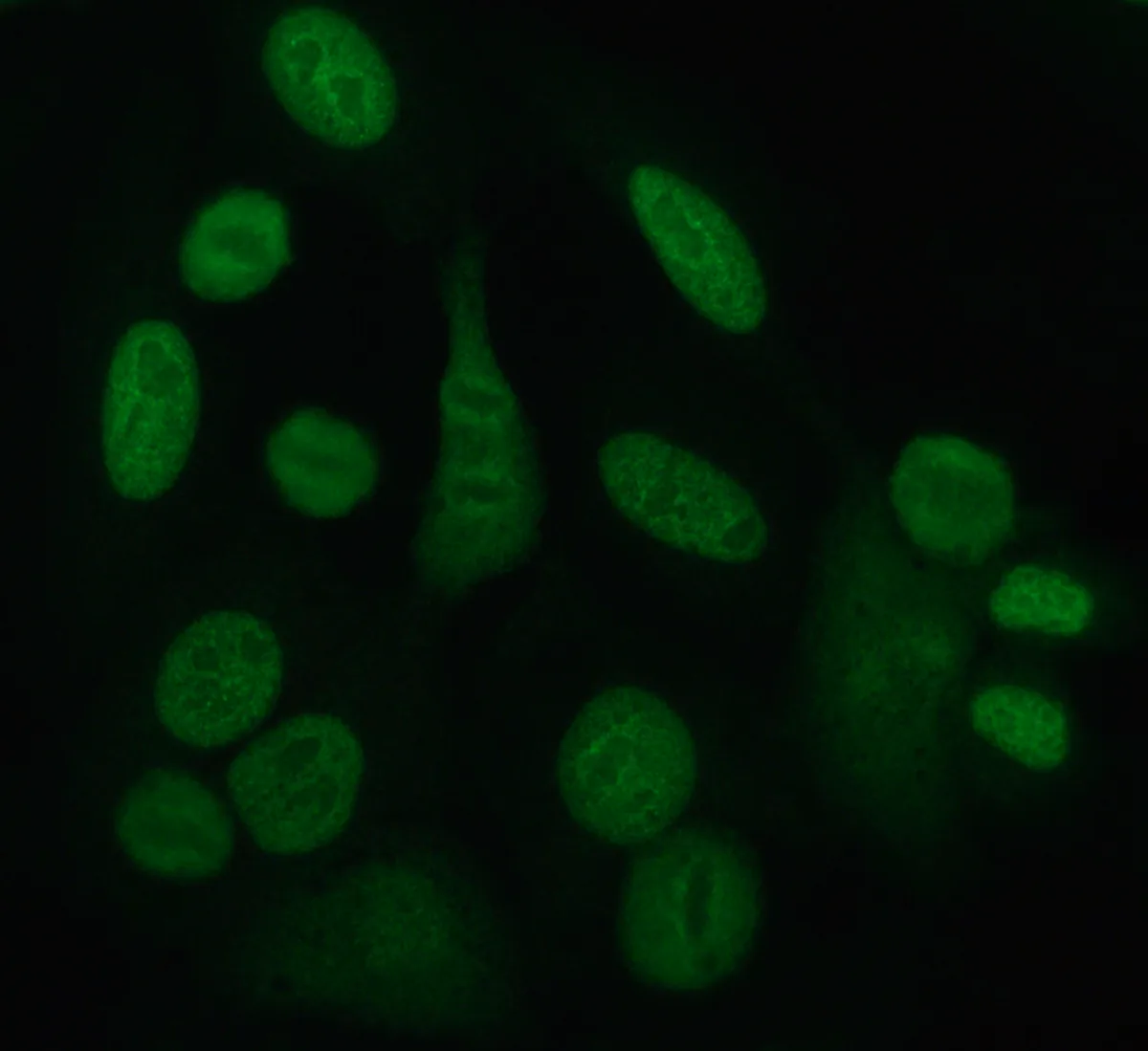
- Autoantibodies: anti-SSA (Ro) (the only antibody included in the classification criteria), anti-SSB (La), ANA; rheumatoid factor in more than 70 %.
- Other laboratory findings: ESR elevated in about 70 %, anemia in 33 %, leukopenia in up to 25 %.
- Labial salivary gland biopsy: multiple large foci of lymphocytes with acinar atrophy; mainly when serology is not conclusive or a major organ is involved.
Keep learning in the app
Further reading (open access)
Cross-references
More topics: Rheumatology & immunology
- Rheumatoid arthritis
- Systemic lupus erythematosus (SLE)
- Granulomatosis with polyangiitis (GPA)
- Polymyalgia rheumatica
- Gout
- Reactive arthritis
- Behçet's disease
- Giant cell arteritis (temporal arteritis)
- Septic arthritis
- Mixed connective tissue disease (Sharp syndrome)
- Psoriatic arthritis
- Uveitis in rheumatic diseases
Note: Learning content for medical education – not a treatment recommendation and no substitute for diagnosis or treatment decisions in individual cases. Treatment and management are deliberately not covered on this page.