Sarcoidosis
Board exam relevance: in 12 of 105 exam reports · rank 20- Synonyms
- Boeck's disease, Löfgren syndrome, Heerfordt syndrome, pulmonary sarcoidosis, sarcoid
- Specialty
- Internal medicine · Pulmonology
- Images
- Diagram 1 · Clinical 1 · Histology 2 · X-ray 1 · CT 1 · Gross specimen 1
- Last updated
- 10/2026 · Dr. Pascal Bafteh
Contents
Images (7)
 Diagram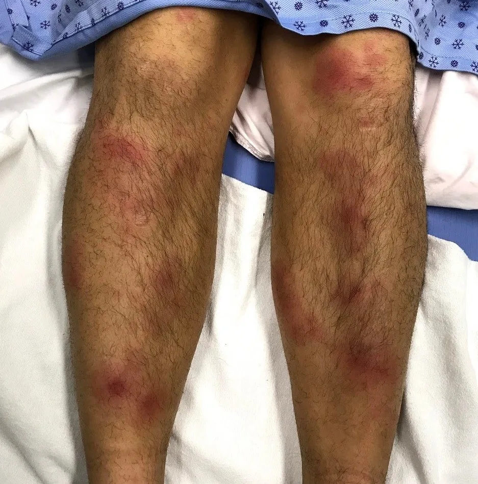
 Histology
 X-ray
X-ray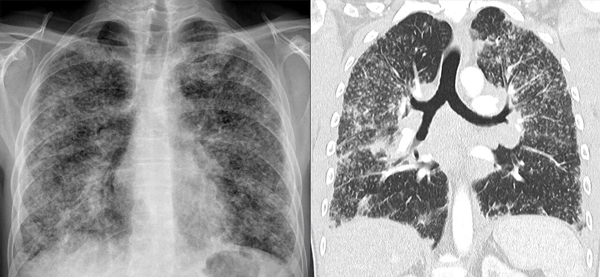 CT
CT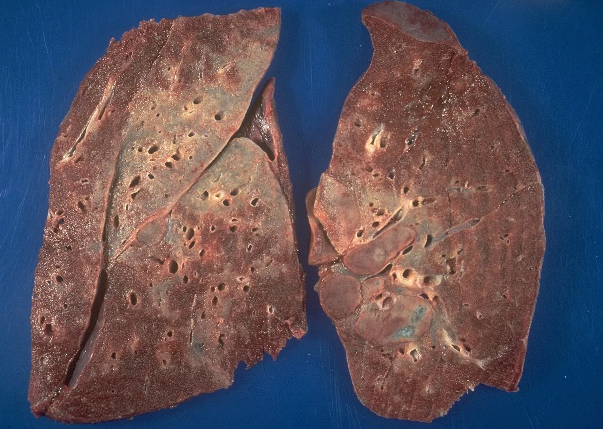 Gross specimen
 Histology
Definition
Sarcoidosis is a granulomatous multisystem disease of unknown cause. Its pathological hallmark is non-caseating epithelioid granulomas, which most often affect the lungs and lymph nodes but can in principle occur in any organ. The disease ranges from asymptomatic incidental findings to chronic courses with organ fibrosis.
Classification
Radiographic stages (Scadding):
| Stage | Chest X-ray finding |
|---|---|
| 0 | normal |
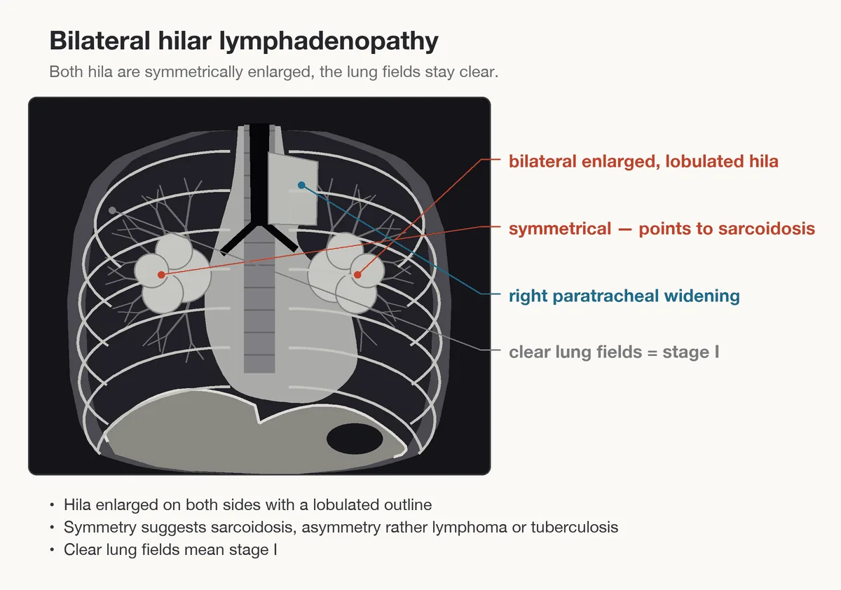
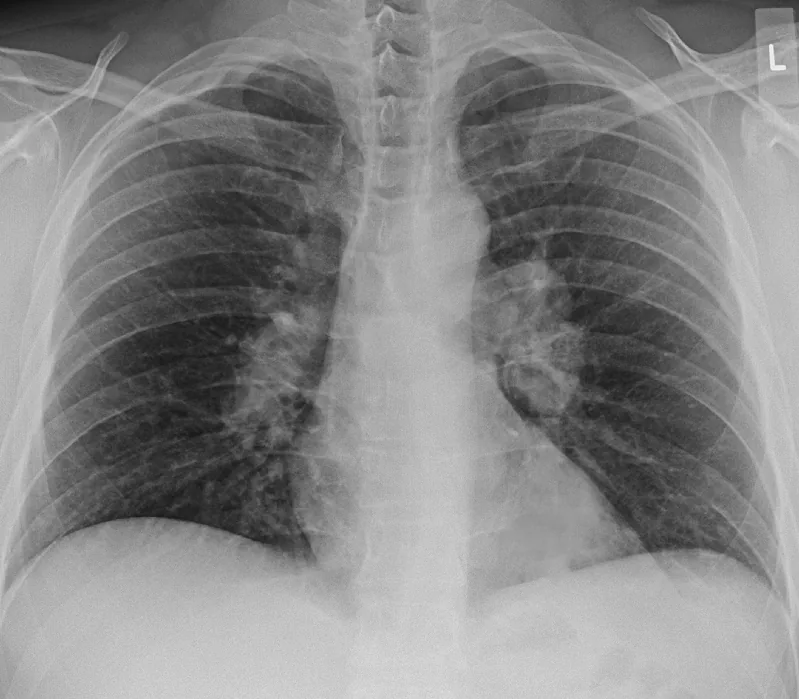
| I | bilateral hilar lymphadenopathy without pulmonary infiltrates |
| II | bilateral hilar lymphadenopathy with pulmonary infiltrates |
| III | pulmonary infiltrates without bilateral hilar lymphadenopathy |
| IV | pulmonary fibrosis |
The stages reflect the extent of lung involvement only roughly and correlate poorly with lung function; a normal chest X-ray does not exclude sarcoidosis, for example with cardiac or neurological involvement.
Clinical special forms:
- Löfgren syndrome: acute form with the triad of acute migratory polyarthritis, erythema nodosum and bilateral hilar lymphadenopathy.
- Heerfordt syndrome (uveoparotid fever): uveitis, bilateral parotid swelling, facial nerve palsy and fever.
Occurrence & epidemiology
Sarcoidosis occurs worldwide, most often between the ages of 20 and 40, occasionally in children and older people. Prevalence is highest in Black Americans and in northern Europeans, especially in Scandinavia. The pattern of manifestations differs: Black patients more often have extrathoracic involvement of the eyes, liver, bone marrow, peripheral lymph nodes and skin, women more often erythema nodosum and eye and nervous system involvement, men and older people more often hypercalcemia. Many cases are asymptomatic and remain undetected.
Aetiopathogenesis
An excessive inflammatory reaction to antigens not yet reliably identified in genetically susceptible people is suspected.
- Proposed triggers: infectious agents (Propionibacterium acnes, mycobacterial antigens), environmental exposures (mold, mildew, musty work environments), occupational exposures (metal work with beryllium, agriculture and pesticides, shipbuilding, firefighting).
- Genetics: higher concordance in identical twins, familial clustering; HLA and non-HLA genes influence risk and course. The haplotype HLA-DRB1*03/DQB1*02 is associated with Löfgren syndrome.
- Smoking: is inversely correlated with sarcoidosis.
Pathophysiology: An unknown antigen triggers a cell-mediated immune reaction with accumulation of T cells and macrophages and release of cytokines such as TNF-α; the cells organize into granulomas. In the lung the granulomas lie along the lymphatics in peribronchial, subpleural and perilobular locations. They may regress, persist or progress to fibrosis. Activated macrophages convert more vitamin D into its active form (1,25-dihydroxyvitamin D); the result is hypercalciuria, hypercalcemia, nephrolithiasis and nephrocalcinosis.
Clinical features
Symptoms depend on the site and extent of organ involvement and change over time. General symptoms such as fatigue, malaise, weakness, loss of weight and low-grade fever are common; sarcoidosis can present as fever of unknown origin.
Organ involvement
- Lungs (over 90 %): often asymptomatic and found incidentally; otherwise breathlessness, dry cough, chest tightness, crackles; rarely pulmonary hypertension.
- Lymph nodes: hilar or mediastinal nodes in most patients, peripheral nodes less often.
- Skin (about 25 %): erythema nodosum on the anterior lower legs (often with arthritis of nearby joints), plaques, macules, papules, subcutaneous nodules, hypo- and hyperpigmentation, lupus pernio (violaceous plaques on the nose, cheeks, ears).
- Eyes (about 12 %): mainly uveitis with blurred vision, photophobia and lacrimation; also conjunctivitis and lacrimal gland involvement.
- Liver (about 12 %): usually asymptomatic with mildly raised liver tests; splenic involvement often asymptomatic.
- Nervous system (under 10 %): cranial nerve palsies, especially facial palsy, involvement of the hypothalamus and pituitary with hypopituitarism or AVP deficiency, meningitis.
- Heart: conduction blocks and arrhythmias, heart failure.
- Other organs: arthritis (ankles, knees, wrists and elbows), myopathy, parotid swelling, bone lesions, renal involvement through hypercalciuria and interstitial nephritis, lymphopenia and anemia.
Histology
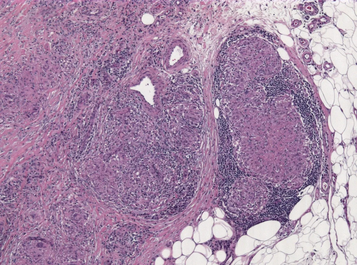
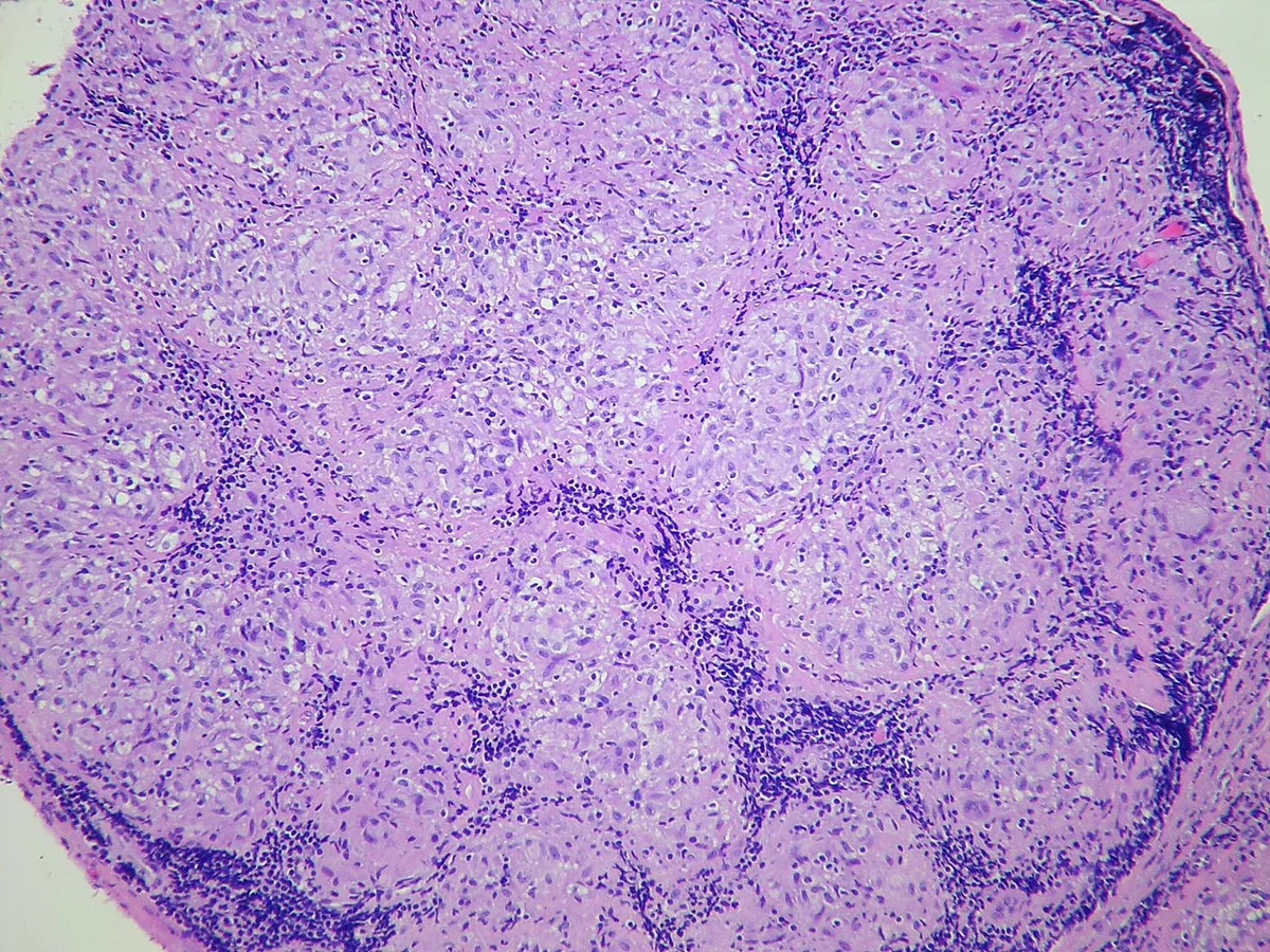
The hallmark is non-caseating granulomas: collections of mononuclear cells and macrophages that differentiate into epithelioid cells and multinucleated giant cells and are surrounded by lymphocytes, plasma cells, fibroblasts and collagen. In the lung they lie along the lymphatics in peribronchiolar, subpleural and perilobular locations and along the bronchovascular bundles.
Diagnosis
The diagnosis is based on a compatible clinical and radiological picture, histological demonstration of non-caseating granulomas and exclusion of other granulomatous diseases (e.g. tuberculosis, fungal infections, berylliosis, hypersensitivity pneumonitis, lymphomas). A typical Löfgren syndrome usually requires no biopsy in the absence of signs of tuberculosis.
Imaging
- Chest X-ray: the most common finding is bilateral hilar lymphadenopathy, often incidental.
- HRCT: more sensitive for hilar and mediastinal lymph nodes and parenchymal changes, predominantly in the upper lobes: thickening of bronchovascular bundles and bronchial walls, beaded interlobular septa, nodules, ground-glass opacities, in advanced stages cysts and traction bronchiectasis.
- PET: detects occult active sites (e.g. bone, muscle, liver, spleen) as a site for biopsy.
Tissue confirmation
- EBUS-guided needle aspiration of mediastinal and hilar lymph nodes, transbronchial lung biopsy or biopsy of easily accessible lesions (skin, peripheral lymph nodes).
- Bronchoalveolar lavage: lymphocytosis (above 15 %) and a CD4/CD8 ratio above 3.5 support the diagnosis in the right context.
Function, laboratory tests and organ screening
- Lung function: often normal in early stages, later restriction and reduced diffusing capacity; 6-minute walk test to detect exercise desaturation.
- Laboratory tests: serum calcium and 24-hour urinary calcium excretion (hypercalciuria even with normal serum calcium), renal and liver tests, blood count. A raised angiotensin-converting enzyme (ACE) may point to sarcoidosis but is nonspecific.
- Search for extrapulmonary involvement: 12-lead ECG, ophthalmological slit-lamp examination; further imaging (e.g. MRI, PET) if cardiac or neurological involvement is suspected.
Keep learning in the app
Further reading (open access)
Cross-references
More topics: Pulmonology
- Community-acquired pneumonia
- Tuberculosis
- Pneumothorax
- Lung cancer (bronchial carcinoma)
- COPD (chronic obstructive pulmonary disease)
- Pleural effusion
- Asthma
- Acute respiratory distress syndrome (ARDS)
- Legionnaires' disease
- Pleural empyema
- Alpha-1 antitrypsin deficiency
- Idiopathic pulmonary fibrosis and interstitial lung disease
Note: Learning content for medical education – not a treatment recommendation and no substitute for diagnosis or treatment decisions in individual cases. Treatment and management are deliberately not covered on this page.