Alpha-1 antitrypsin deficiency
Board exam relevance: in 2 of 105 exam reports · rank 142- Synonyms
- AATD, A1AT deficiency, alpha-1 deficiency, hereditary emphysema, AAT deficiency
- Specialty
- Internal medicine · Pulmonology
- Images
- CT 1 · Histology 2 · Gross specimen 1
- Last updated
- 10/2026 · Dr. Pascal Bafteh
Contents
Images (4)
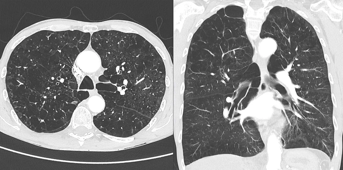 CT
CT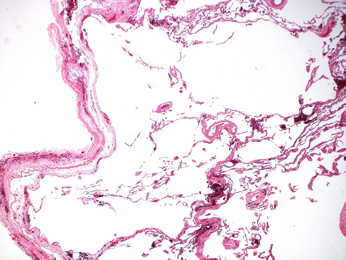 Histology
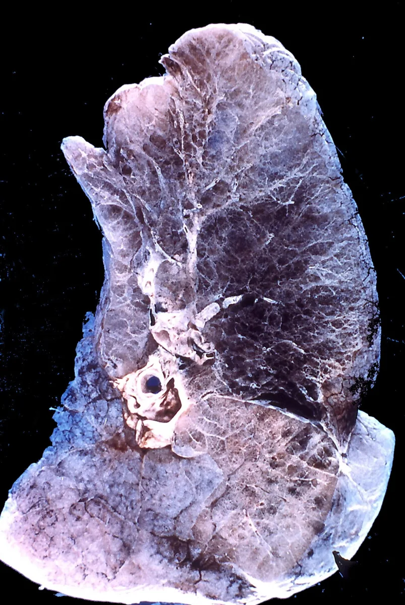 Gross specimen
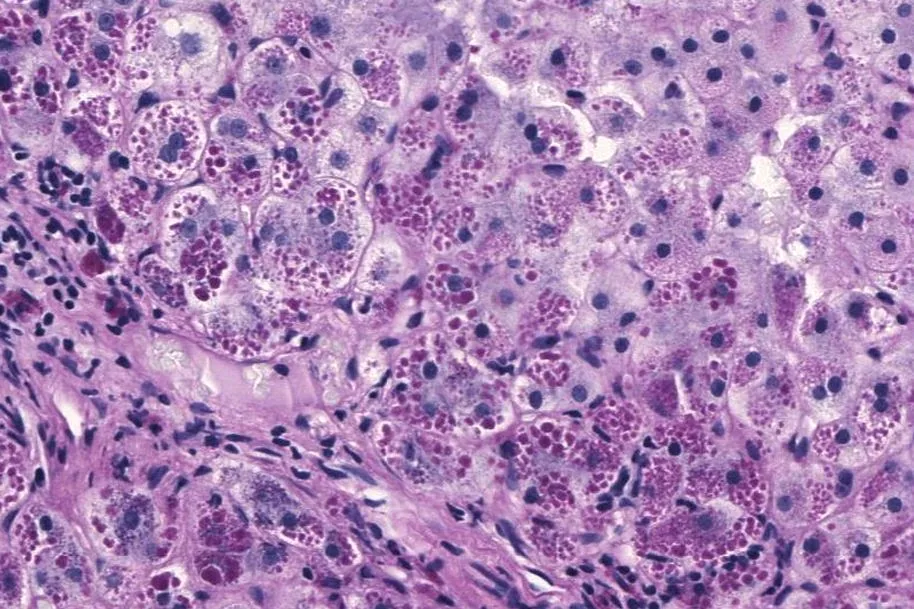 Histology
Definition
Alpha-1 antitrypsin deficiency is a hereditary deficiency of alpha-1 antitrypsin, the main antiprotease of the lung against neutrophil elastase. It leads to increased proteolytic tissue destruction with emphysema in adulthood; accumulation of misfolded molecules in the liver can cause liver disease in children and adults.
Classification
The alleles are expressed codominantly; more than 140 alleles have been described by Pi phenotype.
| Phenotype | Serum level | Emphysema risk |
|---|---|---|
| Pi*MM | 150–350 mg/dL | normal |
| Pi*MZ | 90–210 mg/dL | minimally increased |
| Pi*SS | 100–200 mg/dL | minimally increased |
| Pi*SZ | 75–120 mg/dL | slightly increased |
| Pi*ZZ | 20–45 mg/dL | high |
Over 95 % of people with severe deficiency and emphysema are homozygous for the Z allele (Pi*ZZ). Null alleles (Pi*Z-null, Pi*null-null) result in undetectable serum levels.
Occurrence & epidemiology
The prevalence of the Pi*ZZ genotype in the general population is 1/2,000 to 1/10,000. Mainly people of northern European descent are affected; the Z allele is rare in people of Asian and African descent. About 1–2 % of all COPD cases are attributed to alpha-1 antitrypsin deficiency.
Aetiopathogenesis
Alpha-1 antitrypsin is produced mainly in hepatocytes and monocytes and reaches the lung via the blood, where it neutralizes neutrophil elastase.
- Lung: the deficiency increases neutrophil elastase activity and leads to emphysema, especially in smokers, because cigarette smoke further increases protease activity.
- Liver: certain variants change the conformation of the molecule, which polymerizes and is retained in hepatocytes. This accumulation causes neonatal cholestatic jaundice in 10–15 % of those affected and increases the risk of cirrhosis and hepatocellular carcinoma.
- Other associations: panniculitis, ANCA-positive vasculitis, ulcerative colitis, aneurysms, glomerular diseases.
Clinical features
- Lung: early emphysema with COPD symptoms (breathlessness, cough, wheeze, prolonged expiration); earlier in smokers than in non-smokers, but rare before the age of 25. Severity varies widely with phenotype, smoking status and other factors. Some patients have bronchiectasis.
- Liver: in newborns cholestatic jaundice and hepatomegaly in the first week of life, usually resolving after 2–4 months; in about 20 % the neonatal liver involvement leads to cirrhosis in childhood. About 10 % of patients without childhood liver disease develop cirrhosis in adulthood.
- Skin: panniculitis with indurated, soft, depigmented plaques or nodules, typically on the lower abdomen, buttocks and thighs.
Diagnosis
Suspicion arises in:
- smokers with emphysema before the age of 45
- non-smokers without occupational exposure with emphysema at any age
- a positive family history of emphysema, unexplained cirrhosis or alpha-1 antitrypsin deficiency
- panniculitis, unexplained bronchiectasis or liver disease
- newborns with jaundice or raised liver enzymes
Laboratory tests: a serum level below 80 mg/dL (below 15 µmol/L, radial immunodiffusion) or below 50 mg/dL (below 9 µmol/L, nephelometry) supports the diagnosis; it is confirmed by genotyping or phenotyping. With rare variants, normal levels of a dysfunctional protein may be present.
Keep learning in the app
Further reading (open access)
Cross-references
More topics: Pulmonology
Note: Learning content for medical education – not a treatment recommendation and no substitute for diagnosis or treatment decisions in individual cases. Treatment and management are deliberately not covered on this page.