Tuberculosis
Board exam relevance: in 15 of 105 exam reports · rank 12- Synonyms
- TB, consumption, pulmonary tuberculosis, latent tuberculosis, LTBI
- Specialty
- Internal medicine · Pulmonology
- Images
- X-ray 2 · CT 1 · Blood smear & cytology 1 · Histology 1 · Gross specimen 1 · Diagram 1
- Last updated
- 10/2026 · Dr. Pascal Bafteh
Contents
Images (7)
 X-ray
X-ray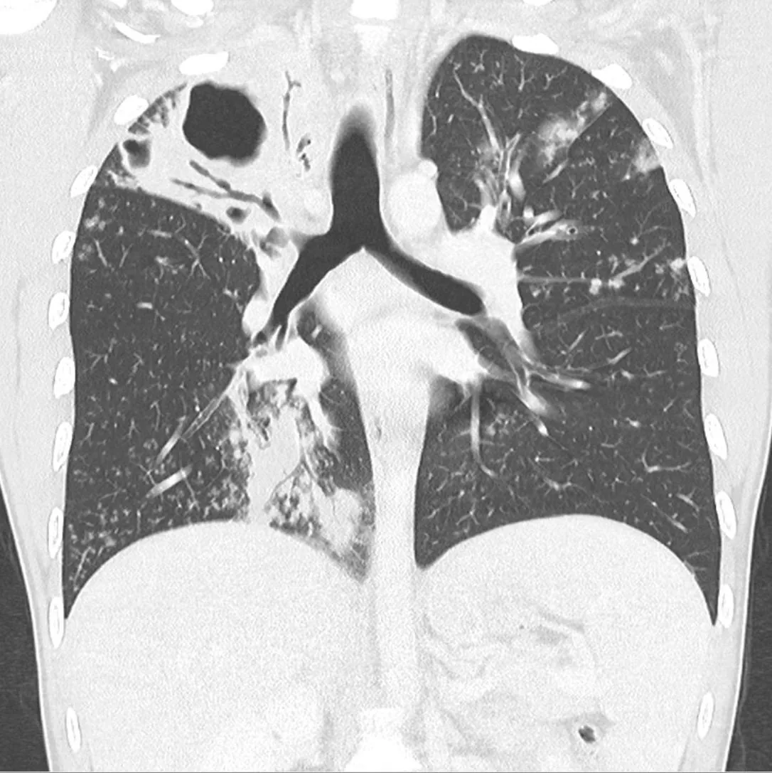 CT
CT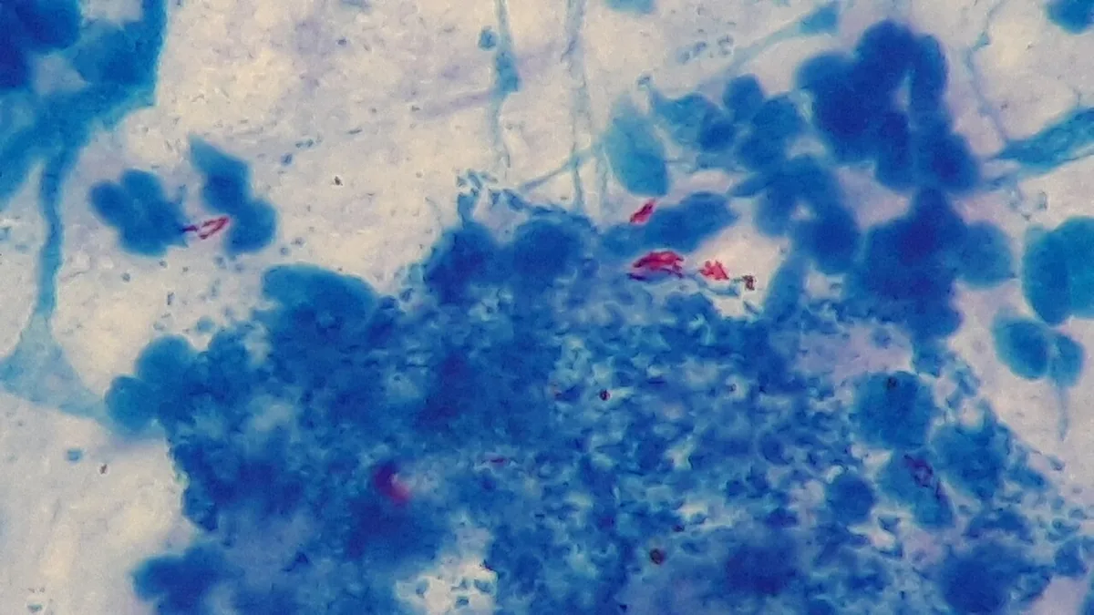 Blood smear & cytology
 Histology
 Gross specimen
 X-ray
X-ray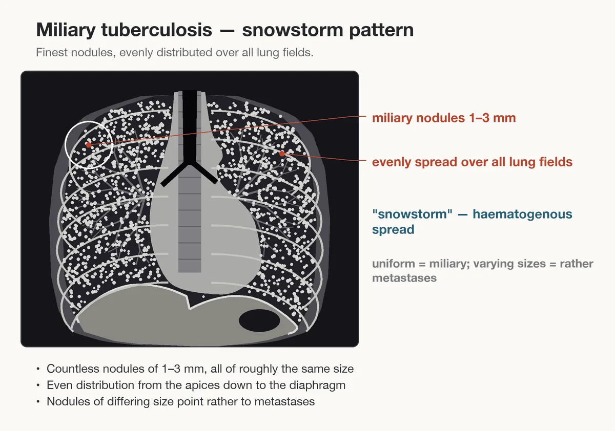 Diagram
Definition
Tuberculosis (TB) is an infectious disease caused by bacteria of the Mycobacterium tuberculosis complex. In about 70 % of cases it manifests as pulmonary tuberculosis, but through hematogenous or lymphatic spread it can affect any organ.
It is distinguished from latent tuberculosis infection (LTBI): viable organizms persist in the body without causing signs of disease; only the immunological reaction can be demonstrated.
Classification
- LTBI and active tuberculosis: LTBI causes no symptoms; active tuberculosis is a disease with clinical, radiological or microbiological findings.
- Primary tuberculosis: disease in temporal connection with the first infection, e.g. as pneumonia with pleural effusion and marked hilar or mediastinal lymph node enlargement.
- Reactivation (post-primary tuberculosis): disease arising from dormant foci, often only after years or decades, preferentially in the apical-posterior lung segments.
- Pulmonary, extrapulmonary and disseminated tuberculosis: involvement of the lung, other organs (e.g. lymph nodes, pleura, urinary tract, bones, joints, spine, meninges) or several organs; miliary tuberculosis in generalized hematogenous spread.
- Infectious ("open") pulmonary tuberculosis: the lesion communicates with the airways; smear-positive cases are particularly infectious, cases detected only by culture or PCR less so.
- MDR-TB: organizms insensitive to the two most important standard drugs.
Occurrence & epidemiology
Worldwide, tuberculosis is one of the most common infectious diseases together with HIV and malaria. According to WHO estimates, about 10.6 million people fall ill each year, almost half a million of them with MDR-TB; about a quarter of the world population is considered infected.
Germany is a low-incidence country: the notification incidence has been below 10 since 2001 and recently around 5 per 100,000 population; about 4,500 cases are registered each year (2023), with a rising trend again since 2022.
Aetiopathogenesis
Pathogen: aerobic, non-motile, slow-growing, acid-fast rods of the M. tuberculosis complex (including M. tuberculosis, M. africanum, M. bovis, M. canetti); the most common human pathogen is M. tuberculosis. Non-tuberculous mycobacteria and M. leprae do not belong to it.
Transmission: almost always airborne by inhaling tiny aerosols containing the organizm (droplet nuclei), expelled mainly by coughing in people with infectious pulmonary tuberculosis. As a rule, several hours of contact in enclosed spaces are required. Children under ten are less often and usually less infectious. Extrapulmonary forms pose no risk of infection in social contacts.
Risk factors: close contact with people with infectious pulmonary tuberculosis, previous tuberculosis, HIV infection, smoking, alcohol and drug dependence, malnutrition, diabetes mellitus, homelessness, imprisonment, poverty and origin from high-prevalence countries. Progression of LTBI is favored by weakened immunity, e.g. uncontrolled HIV infection, older age, congenital immune defects or drug-induced immune suppression such as TNF-α blockade.
Course after infection: On average six to eight weeks pass before a measurable immune reaction develops. In most cases the body eliminates or walls off the organizms. The lifetime risk of disease developing from LTBI is about 5–10 % in immunocompetent adolescents and adults, highest in the first two years. Infants, young children and people with HIV infection and a low CD4 count fall ill much more often (20–40 %) and usually soon after infection.
Pathogenesis
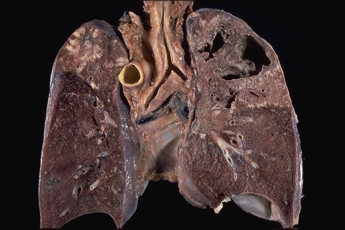
The organizms are taken up by alveolar macrophages and survive intracellularly. The cellular immune reaction gives rise to granulomas; some infected macrophages reach the regional (hilar, mediastinal) lymph nodes and from there spread hematogenously to other organs, especially the well-aerated apical-posterior lung segments. Tissue damage results mainly from a delayed-type hypersensitivity reaction, which can lead to caseous necrosis and cavities.
Residues of the first infection are small consolidations (Ghon focus), together with involved lymph nodes the Ghon complex, when calcified the Ranke complex, and fibronodular scars in the lung apices (Simon foci). In HIV co-infection, granuloma and cavity formation are impaired, with a higher risk of dissemination.
Clinical features
- Cardinal symptom of pulmonary tuberculosis: cough with or without sputum, rarely bloody; occasionally chest pain and breathlessness. Cough lasting more than three weeks raises the possibility of tuberculosis.
- General symptoms (nonspecific): reduced general condition, loss of appetite, unintentional loss of weight, low-grade fever, increased sweating especially at night, fatigue, weakness; failure to thrive in children.
- Oligo- and asymptomatic courses: in a relevant proportion of patients; young children are symptom-free in more than half of cases.
- Hemoptysis: rare, e.g. when a cavity erodes bronchial vessels or an aspergilloma forms in a cavity.
- Extrapulmonary tuberculosis: the most common form is lymph node tuberculosis; also pleurisy, urogenital, bone and joint tuberculosis, spinal involvement (tuberculous spondylitis, Pott disease), tuberculous meningitis.
- Miliary tuberculosis and meningitis: particularly in infants, young children and people with impaired immunity.
- HIV co-infection: often an atypical presentation with more extrapulmonary and disseminated disease; the chest X-ray often shows only a nonspecific infiltrate or is normal.
Histology
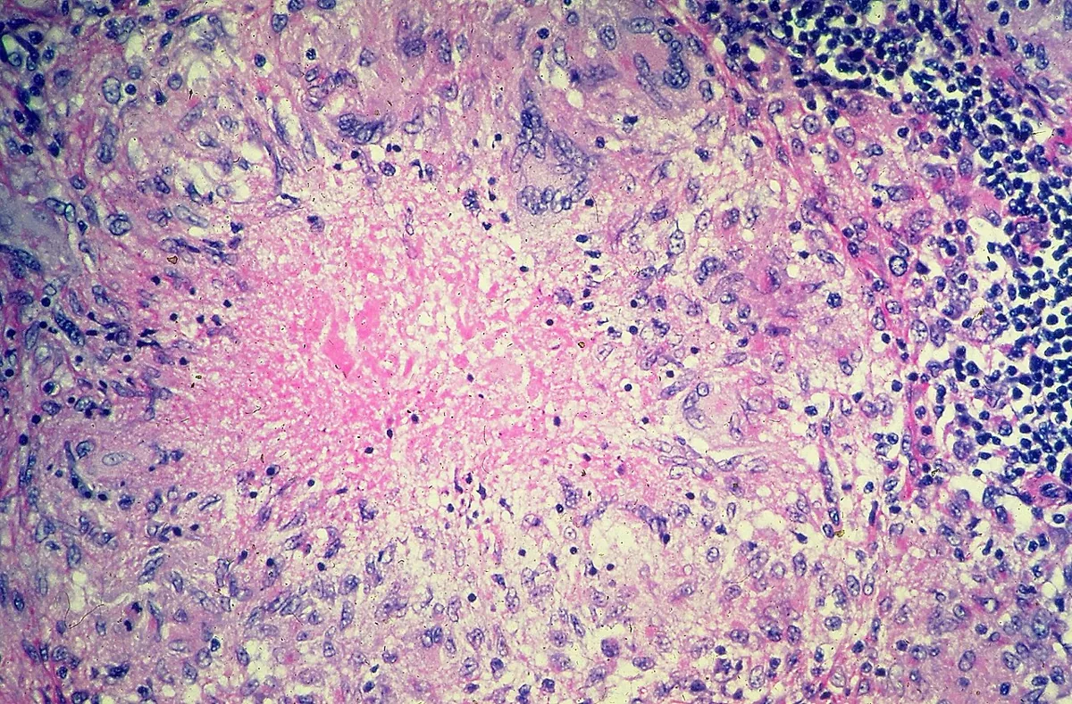
Typical findings are epithelioid granulomas composed of macrophages, epithelioid cells and lymphocytes that enclose the organizms. The granulomas may show central caseous necrosis, in which tubercle bacilli can survive for years. Acid-fast bacilli can be shown with special stains.
Diagnosis
History and imaging
- History: contact with people with infectious pulmonary tuberculosis, occupational exposure, known LTBI or previous tuberculosis, membership of risk groups, origin from or prolonged stay in a high-prevalence country.
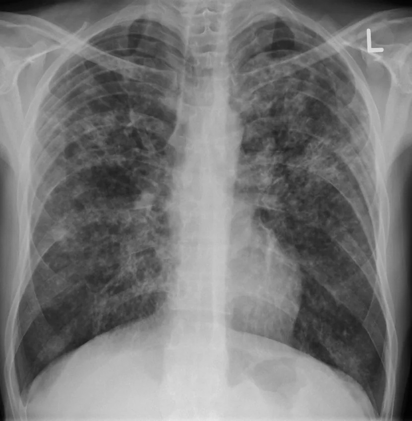
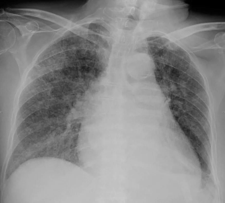
- Chest X-ray: the key investigation when pulmonary tuberculosis is suspected. Most characteristic of active disease in adults is a multinodular infiltrate above or behind the clavicle (reactivation), often with cavities. Infiltrates in the middle and lower zones, especially with pleural effusion, suggest primary tuberculosis in younger people; calcified hilar lymph nodes may be residues of a primary infection.
- Basic laboratory tests: full blood count, liver and renal tests; test for HIV infection, and for hepatitis B and C if risk factors are present.
Microbiology
- Material: For the initial diagnosis three high-quality sputum samples (e.g. early-morning sputum) are examined.
- Microscopy: detection of acid-fast bacilli, preferably by fluorescence microscopy (superior to Ziehl-Neelsen staining). Positive only from about 10⁴ organizms per mL of sputum; it cannot distinguish tubercle bacilli from non-tuberculous mycobacteria.
- Nucleic acid amplification (e.g. PCR): rapid detection of the M. tuberculosis complex from about 10–100 colony-forming units per mL and thus considerably more sensitive than microscopy. In smear-positive samples sensitivity and specificity are close to 100 %; in smear-negative, culture-positive respiratory samples at most about 80 %, so a negative test does not exclude tuberculosis. Depending on the method, common mutations indicating insensitivity to the most important standard drugs are detected at the same time.
- Culture: the most sensitive method and conclusive; in liquid culture systems usually positive after one to three weeks, while a negative result generally requires eight weeks of incubation. The first isolate undergoes species identification and phenotypic and genotypic susceptibility testing.
Immunological tests
Interferon-gamma release assays (IGRA) and the tuberculin skin test (TST) show whether the immune system has been exposed to M. tuberculosis. They become positive at the earliest six to eight weeks after infection. The TST is less specific, as it can be falsely positive after BCG exposure or infection with certain non-tuberculous mycobacteria. Neither test distinguishes between LTBI, active disease and previous disease; they serve mainly to demonstrate or exclude LTBI. False-negative results are possible with weakened immunity or severe disease (e.g. miliary tuberculosis).
Keep learning in the app
Further reading (open access)
Cross-references
More topics: Pulmonology
- Community-acquired pneumonia
- Pneumothorax
- Sarcoidosis
- Lung cancer (bronchial carcinoma)
- COPD (chronic obstructive pulmonary disease)
- Pleural effusion
- Asthma
- Acute respiratory distress syndrome (ARDS)
- Legionnaires' disease
- Pleural empyema
- Alpha-1 antitrypsin deficiency
- Idiopathic pulmonary fibrosis and interstitial lung disease
Note: Learning content for medical education – not a treatment recommendation and no substitute for diagnosis or treatment decisions in individual cases. Treatment and management are deliberately not covered on this page.