Lung cancer (bronchial carcinoma)
Board exam relevance: in 11 of 105 exam reports · rank 24- Synonyms
- bronchial carcinoma, bronchogenic carcinoma, NSCLC, SCLC, small cell lung cancer, non-small cell lung cancer
- Specialty
- Internal medicine · Pulmonology
- Images
- Diagram 1 · X-ray 1 · CT 1 · Gross specimen 1 · Histology 2
- Last updated
- 10/2026 · Dr. Pascal Bafteh
Contents
Images (6)
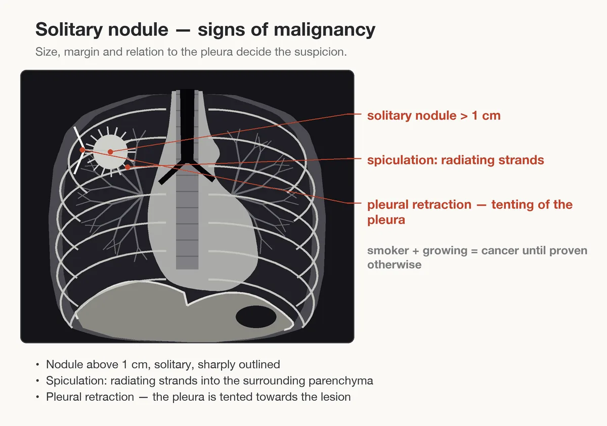 Diagram
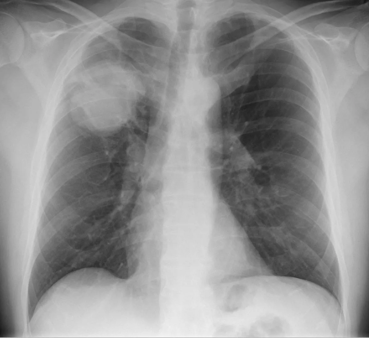 X-ray
X-ray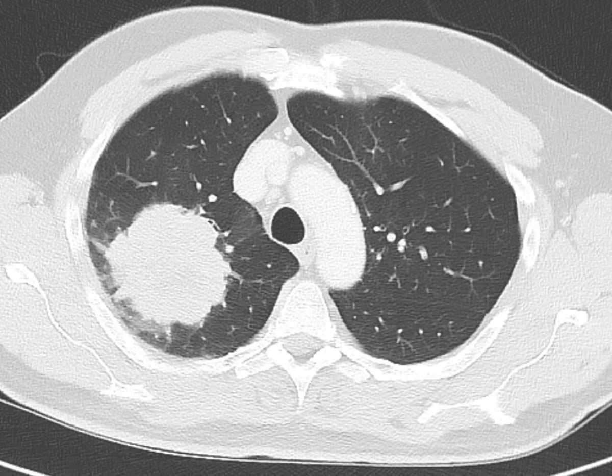 CT
CT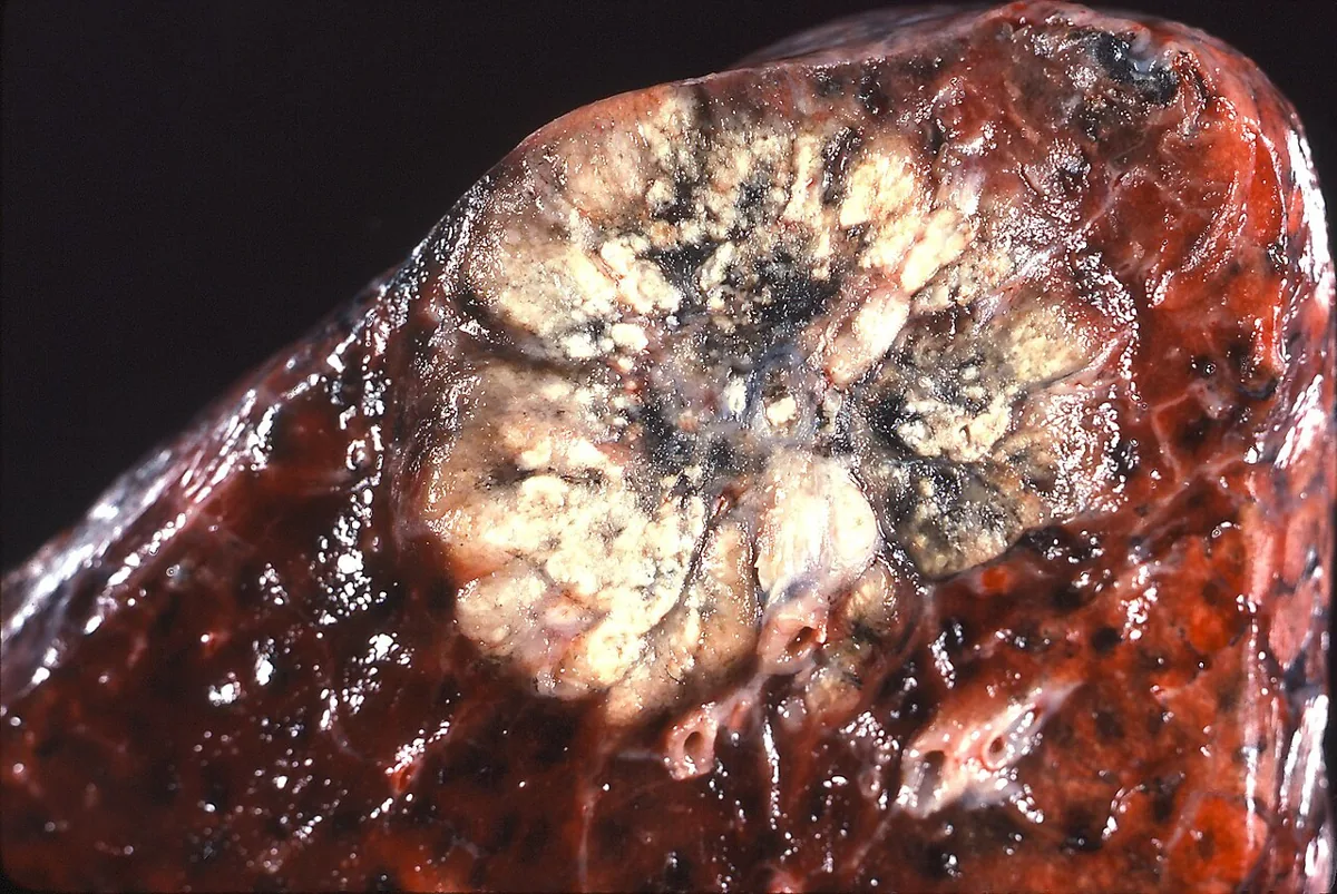 Gross specimen
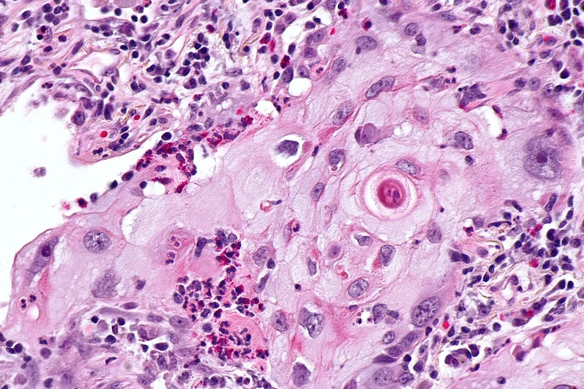 Histology
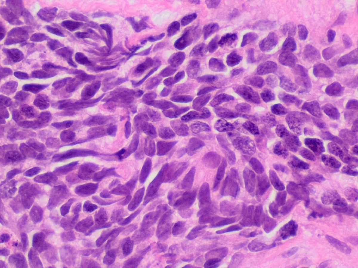 Histology
Definition
Lung cancer (bronchial carcinoma) is a malignant epithelial tumor arising from the airways or the lung parenchyma. Histologically, two main groups are distinguished: small cell (SCLC) and non-small cell lung cancer (NSCLC).
Classification
| Type | Share | Typical location |
|---|---|---|
| Small cell carcinoma (SCLC) | 13–15 % | submucosal in the airways, perihilar mass |
| Adenocarcinoma | 40 % | peripheral nodule or mass |
| Squamous cell carcinoma | 25–30 % | central, endobronchial |
| Large cell carcinoma | 10–15 % | peripheral nodule or mass |
TNM classification
Spread is described by the TNM system (currently 9th edition):
- T1: tumor ≤ 3 cm (T1a ≤ 1 cm, T1b > 1–2 cm, T1c > 2–3 cm).
- T2: tumor > 3–5 cm or with certain features such as invasion of the visceral pleura.
- T3: tumor > 5–7 cm or invasion e.g. of the chest wall, pericardium, phrenic nerve; separate tumor nodule in the same lobe.
- T4: tumor > 7 cm or invasion of the diaphragm, mediastinum, heart, great vessels, trachea, recurrent laryngeal nerve, esophagus, vertebral body or carina.
- N1: ipsilateral peribronchial and/or hilar or intrapulmonary lymph nodes. N2: ipsilateral mediastinal and/or subcarinal lymph nodes, subdivided into N2a (single station) and N2b (multiple stations). N3: contralateral mediastinal or hilar, scalene or supraclavicular lymph nodes.
- M1a: tumor nodules in the contralateral lung, pleural or pericardial nodules, malignant pleural or pericardial effusion. M1b: single extrathoracic metastasis in one organ. M1c1/M1c2: multiple extrathoracic metastases in one or several organ systems.
SCLC is also divided into limited stage (confined to one hemithorax including ipsilateral lymph nodes, without malignant pleural or pericardial effusion) and extensive stage. Only about one in three patients with SCLC has limited-stage disease at diagnosis.
Occurrence & epidemiology
Lung cancer is the leading cause of cancer death worldwide. About 2.5 million new cases were estimated for 2022, around 12 % of all newly diagnosed cancers. Men are affected more often. Differences between regions and sexes are partly explained by differences in smoking prevalence.
Aetiopathogenesis
- Cigarette smoking: the most important cause, responsible for about 85 % of cases; active smoking (including cigars) and passive smoking. The risk increases with the intensity and duration of smoking and falls after quitting but never returns to the level of never-smokers.
- Never- or light smokers: about 15–20 % of patients; oncogenic driver mutations are often found in them.
- Radon: chronic residential exposure increases the risk and is considered the second most common cause in the USA.
- Occupational and environmental carcinogens: asbestos, arsenic, cadmium, chromium, nickel, chloromethyl ethers, polycyclic aromatic hydrocarbons, coke oven emissions, air pollution, smoke from open cooking and heating fires.
- Lung diseases: COPD, alpha-1 antitrypsin deficiency, pulmonary fibrosis and scarring, e.g. after tuberculosis.
- Family history and previous cancer.
Pathogenesis: Long-term carcinogen exposure leads, through stepwise accumulation of genetic and epigenetic changes in the respiratory epithelium, to malignant transformation (field cancerization). Important are activating alterations in oncogenes such as KRAS, EGFR, BRAF, MET, ERBB2 (HER2), ALK fusions (EML4::ALK), ROS1 and NTRK, as well as loss of tumor suppressor genes such as TP53. SCLC is characterized by an almost universal loss of function of TP53 and RB1.
Clinical features
About 25 % of patients are symptom-free at diagnosis; the tumor is then found incidentally on imaging. In SCLC, 70–80 % of patients already have metastatic disease at diagnosis.
Symptoms
- Local tumor: usually a dry, persistent cough; breathlessness from airway narrowing, post-obstructive atelectasis or pneumonia (with fever); hemoptysis, the first symptom in about 30 %; wheeze with bronchial location; dull chest pain in about 18 %.
- Regional spread: pleural effusion with pleuritic pain and breathlessness; hoarseness from recurrent laryngeal nerve involvement; raised hemidiaphragm from phrenic nerve palsy; superior vena cava syndrome with head fullness, swelling of the face and arms, dilated neck and chest wall veins and plethora; dysphagia; pericardial effusion.
- Pancoast tumor: apical tumor (usually NSCLC) invading the brachial plexus, pleura or ribs: pain in the shoulder and arm, weakness and atrophy of the hand muscles, often Horner syndrome (ptosis, miosis, anhidrosis).
- Distant metastases: brain (confusion, seizures, paralysis), bone (pain, pathological fractures), liver (pain, jaundice), adrenal glands.
- General symptoms: loss of appetite, cachexia, loss of weight, fatigue.
Paraneoplastic syndromes
- Hypercalcemia: in squamous cell carcinoma through production of parathyroid hormone-related protein (PTHrP) or extensive bone metastases
- Syndrome of inappropriate ADH secretion (SIADH): in SCLC
- Ectopic Cushing syndrome due to ACTH production: mainly in SCLC
- Lambert-Eaton myasthenic syndrome and other neurological syndromes (neuropathies, encephalitis, cerebellar syndromes): mainly in SCLC
- Digital clubbing with or without hypertrophic pulmonary osteoarthropathy: most common in NSCLC
- Hypercoagulability with migratory thrombophlebitis (Trousseau syndrome)
Histology
- Adenocarcinoma: the most common subtype, especially in never-smokers; variants are adenocarcinoma in situ, minimally invasive and invasive adenocarcinoma with acinar, papillary, micropapillary, lepidic or solid growth patterns.
- Squamous cell carcinoma: keratinization and/or intercellular bridges or immunohistochemical markers of squamous differentiation.
- Large cell carcinoma: undifferentiated NSCLC without features of adenocarcinoma or squamous cell carcinoma; a diagnosis of exclusion after immunohistochemical exclusion of other subtypes.
- Small cell carcinoma: extremely aggressive and fast-growing, almost always in smokers; typically submucosal in the airways or as a perihilar mass.
Diagnosis
Imaging and staging
- Chest X-ray: often the first investigation; suggestive findings are a nodule or mass, enlarged hilum, widened mediastinum, tracheobronchial narrowing, atelectasis, persistent infiltrates, cavities, unexplained pleural thickening or pleural effusion.
- Chest CT: characterization of the lesion and the basis of staging.
- PET-CT: shows metabolically active foci in pulmonary nodules, hilar and mediastinal lymph nodes and distant metastases; very sensitive with a high negative predictive value but limited specificity (false positives e.g. with inflammation).
- Brain MRI (or contrast-enhanced head CT) to detect brain metastases; chest MRI for Pancoast tumors.
Tissue diagnosis
The diagnosis requires pathological confirmation by cytology or tissue sampling, which also allows histological and molecular analysis.
- Cytology of sputum or pleural effusion: the least invasive approach; a malignant pleural effusion indicates advanced disease.
- Bronchoscopy with endobronchial ultrasound (EBUS): the most common procedure for diagnosis and mediastinal lymph node staging; washing, brushing and biopsy of endobronchial lesions and needle aspiration of paratracheal, subcarinal, mediastinal and hilar lymph nodes.
- Percutaneous CT-guided core biopsy: for peripheral lesions and extrathoracic sites; risk of pneumothorax about 25 %, especially in emphysema.
- Molecular pathology: testing for oncogenic driver alterations (e.g. EGFR, ALK, ROS1, BRAF, KRAS, MET, NTRK, HER2) and immunohistochemical assessment of PD-L1 expression.
Keep learning in the app
Further reading (open access)
Cross-references
More topics: Pulmonology
Note: Learning content for medical education – not a treatment recommendation and no substitute for diagnosis or treatment decisions in individual cases. Treatment and management are deliberately not covered on this page.