Acute respiratory distress syndrome (ARDS)
Board exam relevance: in 3 of 105 exam reports · rank 111- Synonyms
- acute lung injury, shock lung, adult respiratory distress syndrome, ALI
- Specialty
- Internal medicine · Pulmonology
- Images
- X-ray 1 · Histology 1 · Gross specimen 1
- Last updated
- 10/2026 · Dr. Pascal Bafteh
Contents
Images (3)
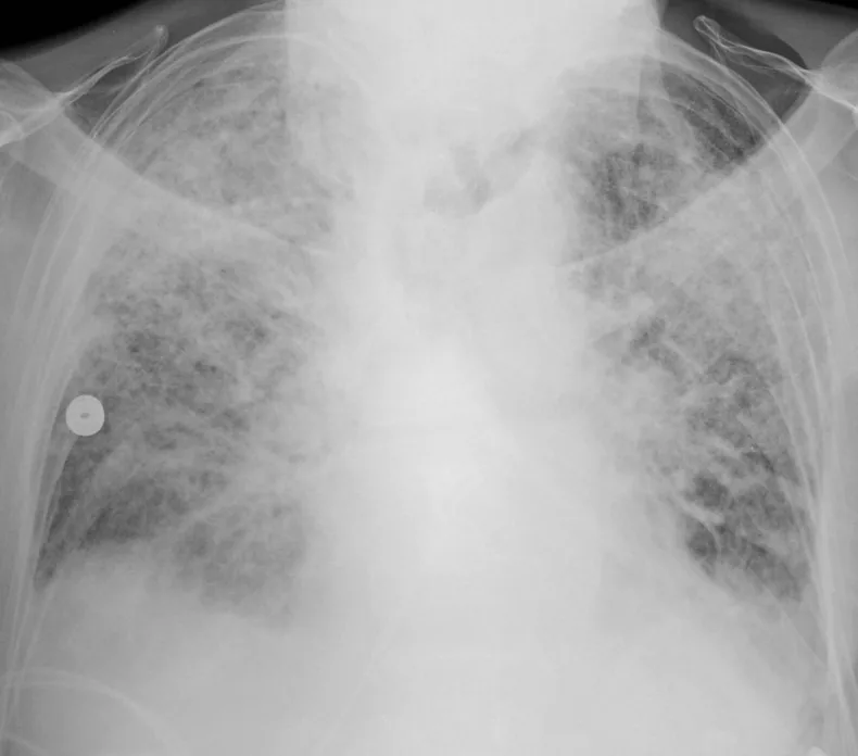 X-ray
X-ray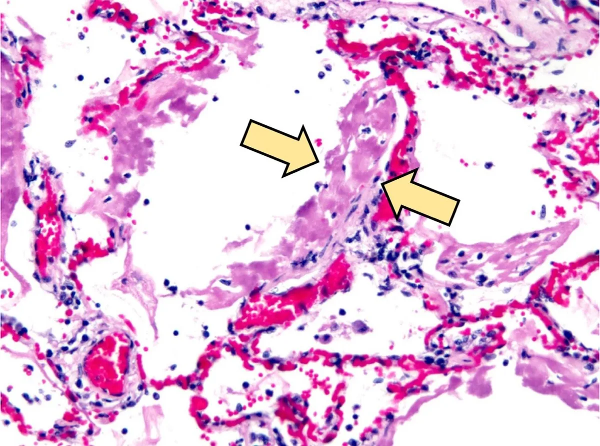 Histology
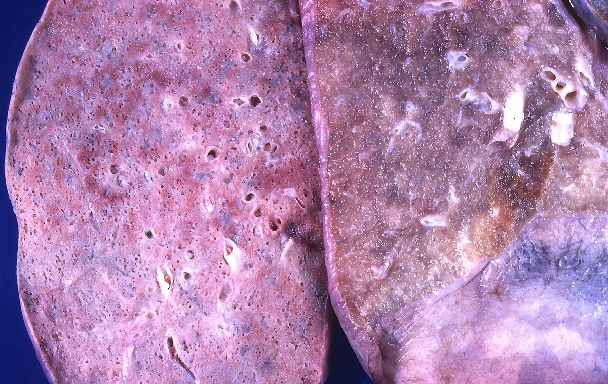 Gross specimen
Definition
Acute respiratory distress syndrome (ARDS) is a diffuse inflammatory lung injury and a cause of acute hypoxemic respiratory failure. Increased permeability of the alveolar-capillary barrier leads to non-cardiogenic pulmonary edema with severe hypoxemia.
Classification
Berlin definition:
- Timing: onset within one week of a known insult or of new or worsening respiratory symptoms.
- Imaging (X-ray or CT): bilateral opacities not fully explained by effusions, lobar or lung collapse or nodules.
- Origin of edema: respiratory failure not fully explained by cardiac failure or fluid overload.
| Severity | PaO₂/FiO₂ (with PEEP ≥ 5 cmH₂O) |
|---|---|
| mild | > 200 to ≤ 300 mmHg |
| moderate | > 100 to ≤ 200 mmHg |
| severe | ≤ 100 mmHg |
The mild grade corresponds to the former category "acute lung injury" (ALI). A newer global definition extends the criteria: ultrasound to demonstrate bilateral opacities, inclusion of patients on high-flow oxygen (at least 30 L/min) and a ratio of oxygen saturation to FiO₂ of ≤ 315 provided saturation is ≤ 97 %.
Aetiopathogenesis
Sepsis and pneumonia account for about 60 % of ARDS cases.
- Direct lung injury – common: aspiration of gastric acid, pneumonia.
- Direct lung injury – less common: diffuse alveolar hemorrhage, lung contusion, drowning, fat embolism, inhalation of irritant gases, amniotic fluid embolism.
- Indirect lung injury – common: sepsis, trauma with prolonged hypovolemic shock.
- Indirect lung injury – less common: pancreatitis, burns, poisoning (e.g. cocaine, opioids, tricyclic antidepressants), neurogenic pulmonary edema after stroke, seizure or head injury, pre-eclampsia.
Pathophysiology: Pulmonary or systemic inflammation releases cytokines that activate alveolar macrophages and recruit neutrophils into the lung. These release leukotrienes, oxidants, platelet-activating factor and proteases and damage the capillary endothelium and alveolar epithelium. Edema fluid, protein and cell debris enter the alveoli and interstitium, destroy surfactant and cause collapse of airspaces, mainly in the dependent lung regions; the results are shunt, severe hypoxemia and pulmonary hypertension. This exudative phase is followed by a fibroproliferative phase with proliferation of the alveolar epithelium and fibrosis. Because flooded or collapsed alveoli take up no gas, the hypoxemia improves little with a higher inspired oxygen fraction.
Clinical features
- Breathlessness, restlessness and anxiety, tachypnea, tachycardia, cyanosis, increased sweating
- Confusion up to impaired consciousness; later arrhythmias and coma
- Diffuse inspiratory crackles, sometimes more marked at the bases
- Signs of the underlying condition, e.g. sepsis, pneumonia or pancreatitis
Diagnosis
- Pulse oximetry and arterial blood gas analysis: detection and severity of hypoxemia (PaO₂/FiO₂ ratio).
- Chest X-ray: bilateral opacities; early on, hypoxemia may precede radiographic changes.
- Search for the cause: pulmonary and extrapulmonary triggers (e.g. pneumonia, sepsis, pancreatitis, trauma, aspiration).
- Distinction from cardiogenic pulmonary edema: in ARDS, respiratory failure is not fully explained by cardiac failure or fluid overload; cardiac function is assessed for this purpose, e.g. by echocardiography.
Keep learning in the app
Further reading (open access)
Cross-references
More topics: Pulmonology
Note: Learning content for medical education – not a treatment recommendation and no substitute for diagnosis or treatment decisions in individual cases. Treatment and management are deliberately not covered on this page.