Idiopathic pulmonary fibrosis and interstitial lung disease
Board exam relevance: in 2 of 105 exam reports · rank 142- Synonyms
- pulmonary fibrosis, IPF, ILD, lung scarring, diffuse parenchymal lung disease, UIP
- Specialty
- Internal medicine · Pulmonology
- Images
- CT 1 · Histology 1 · Gross specimen 1
- Last updated
- 10/2026 · Dr. Pascal Bafteh
Contents
Images (3)
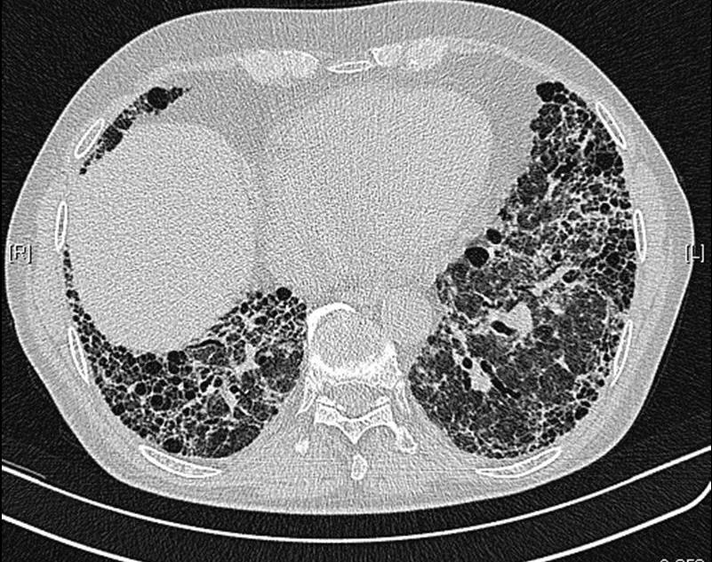 CT
CT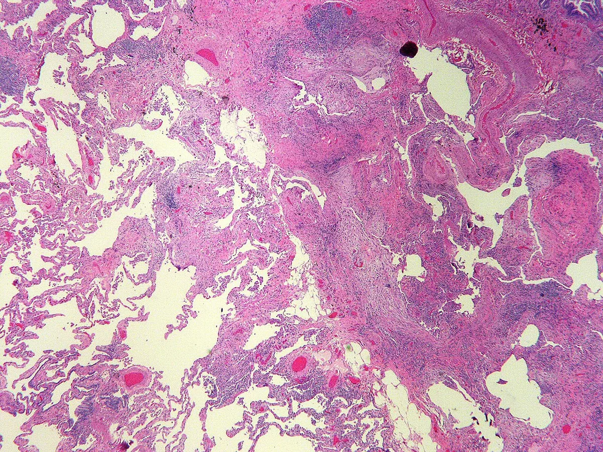 Histology
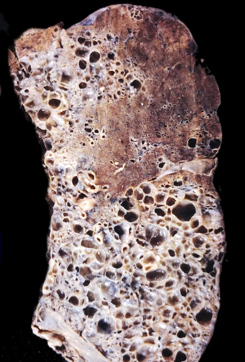 Gross specimen
Definition
Interstitial lung diseases (ILD, diffuse parenchymal lung diseases) are a heterogeneous group of disorders whose pathological processes primarily affect the lung interstitium and spread to the alveoli, bronchioles and bronchi. Characteristic features are diffuse thickening of the alveolar septa, fibroblast proliferation and collagen deposition, progressing to pulmonary fibrosis if unchecked.
Idiopathic pulmonary fibrosis (IPF) is a fibrosing disease of unknown cause with the histological pattern of usual interstitial pneumonia (UIP); it accounts for most cases of idiopathic interstitial pneumonia.
Classification
Among others, acute and chronic, granulomatous and non-granulomatous forms and diseases of known and unknown cause are distinguished:
- Systemic rheumatic diseases: e.g. rheumatoid arthritis, systemic sclerosis, Sjögren syndrome, systemic lupus erythematosus, myositis (especially antisynthetase syndrome), mixed connective tissue disease.
- Iatrogenic and toxic: numerous drugs, including cytotoxic drugs and antiarrhythmics, and illicit substances such as cocaine.
- Occupational and environmental exposures: inorganic dusts (asbestosis, silicosis, berylliosis, coal workers' pneumoconiosis, hard metals) and organic antigens (hypersensitivity pneumonitis).
- Idiopathic interstitial pneumonias: IPF, nonspecific interstitial pneumonia, cryptogenic organizing pneumonia, desquamative interstitial pneumonia, acute interstitial pneumonia, lymphoid interstitial pneumonia, pleuroparenchymal fibroelastosis.
- Others: sarcoidosis, vasculitides, eosinophilic pneumonias, pulmonary Langerhans cell histiocytosis, lymphangioleiomyomatosis, pulmonary alveolar proteinosis, genetic syndromes such as Hermansky-Pudlak syndrome.
Occurrence & epidemiology
The prevalence of interstitial lung disease in Europe and the USA is estimated at about 75 per 100,000 people, with a slight male predominance; older age, especially over 70, is a risk factor. IPF affects people over 50, men about twice as often as women, with incidence rising markedly with each further decade of life.
Aetiopathogenesis
In IPF, repeated small injuries damage alveolar epithelial cells that are already prone to senescence. Environmental factors (above all current or former smoking), genetic factors and other unknown influences lead to epithelial dysfunction and excessive fibroproliferation. A genetic predisposition through variants of the mucin gene MUC5B is found in about 30–35 % of cases. Changes in the lung microbiome and occupational exposure to fumes are also discussed.
Clinical features
- Exertional breathlessness and non-productive cough increasing over months to years in almost all patients with IPF; general symptoms such as low-grade fever and myalgia are rare
- Fine, dry inspiratory crackles at both lung bases ("velcro" crackles)
- Digital clubbing in about half of patients, sometimes acrocyanosis
- Later signs of pulmonary hypertension and right ventricular dysfunction
Histology
The UIP pattern shows subpleural fibrosis with fibroblast foci and dense scarring alternating with areas of normal lung parenchyma (heterogeneity), diffuse interstitial inflammation with lymphocytes, plasma cells and histiocytes, and cystic remodeling (honeycomb lung) that increases as the disease progresses.
Diagnosis
- Chest X-ray: diffuse reticular markings in the lower and peripheral lung zones; in advanced fibrosis small cystic lesions (honeycombing) and traction bronchiectasis.
- HRCT: required for the diagnosis of IPF; typical findings are patchy, subpleural reticular changes with thickened interlobular septa, subpleural honeycombing and traction bronchiectasis. Extensive ground-glass opacities, nodules or a mosaic pattern with air trapping suggest another diagnosis.
- Lung function: restrictive pattern; diffusing capacity is reduced in almost all patients with IPF; exercise hypoxemia.
- Laboratory tests: play a minor role except to exclude systemic rheumatic diseases.
- Lung biopsy: if the diagnosis remains uncertain after the initial evaluation.
HRCT patterns UIP and NSIP
The international guideline (ATS/ERS/JRS/ALAT) classifies HRCT findings into four categories: UIP pattern, probable UIP pattern, indeterminate for UIP, and findings suggesting an alternative diagnosis.
- UIP pattern: subpleural and basal predominant, often heterogeneous (normal lung interspersed with fibrosis); honeycombing with or without traction bronchiectasis or bronchiolectasis, usually with a reticular pattern and mild ground-glass opacities.
- Probable UIP pattern: also subpleural and basal predominant; reticular pattern with traction bronchiectasis or bronchiolectasis, without subpleural sparing.
- Suggestive of NSIP: peribronchovascular predominant changes with subpleural sparing.
Keep learning in the app
Further reading (open access)
- MSD Manual Profi-Ausgabe: Interstitielle Lungenerkrankungen
- MSD Manual Profi-Ausgabe: Idiopathische pulmonale Fibrose
- StatPearls: Idiopathic Pulmonary Fibrosis
- Raghu G et al.: Idiopathic Pulmonary Fibrosis (an Update) and Progressive Pulmonary Fibrosis in Adults – ATS/ERS/JRS/ALAT-Leitlinie 2022 (PMC9851481)
Cross-references
Note: Learning content for medical education – not a treatment recommendation and no substitute for diagnosis or treatment decisions in individual cases. Treatment and management are deliberately not covered on this page.