Pneumothorax
Board exam relevance: in 12 of 105 exam reports · rank 20- Synonyms
- PTX, collapsed lung, lung collapse, tension pneumothorax, spontaneous pneumothorax
- Specialty
- Internal medicine · Pulmonology
- Images
- X-ray 2 · Diagram 1 · CT 1
- Last updated
- 10/2026 · Dr. Pascal Bafteh
Contents
Images (4)
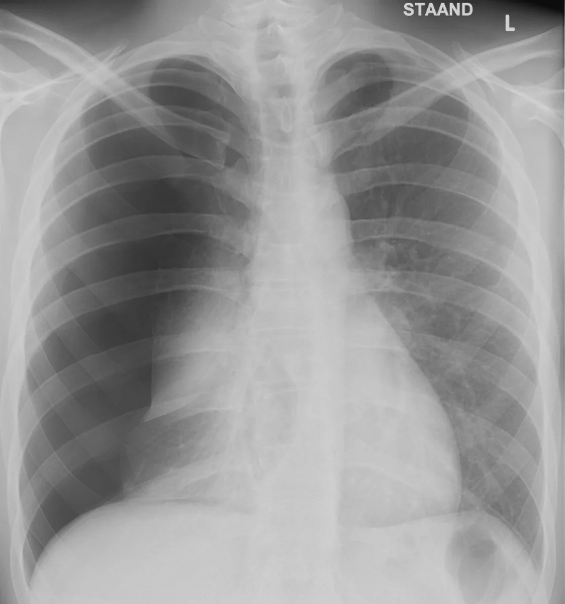 X-ray
X-ray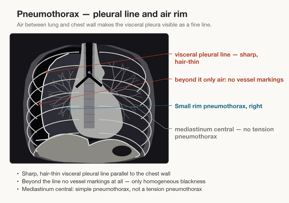 Diagram
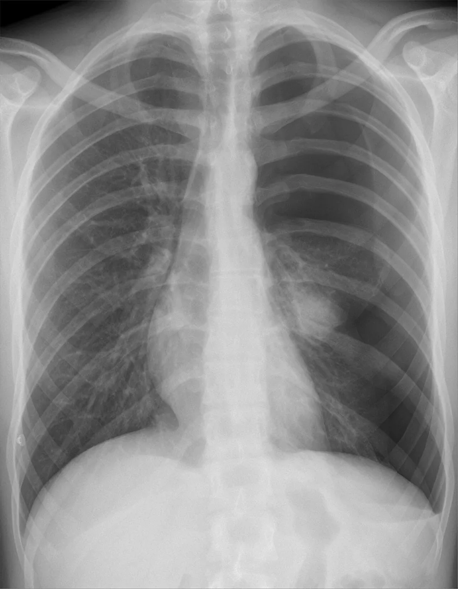 X-ray
X-ray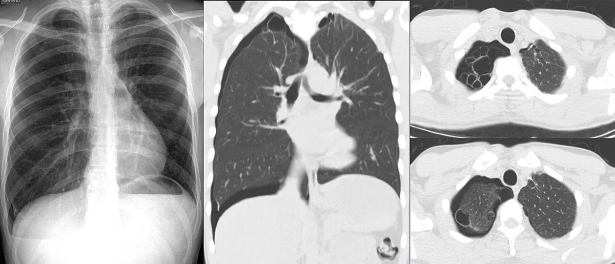 CT
CTDefinition
A pneumothorax is an accumulation of air in the pleural space that causes partial or complete collapse of the lung. It can occur spontaneously or result from an injury or a medical procedure.
Classification
- Primary spontaneous pneumothorax: without underlying lung disease.
- Secondary spontaneous pneumothorax: with pre-existing lung disease; more serious because the underlying disease already reduces lung volume.
- Catamenial pneumothorax: a rare secondary form within 48 hours of the onset of menstruation due to intrathoracic endometriosis.
- Traumatic pneumothorax: a common consequence of penetrating or blunt chest injuries.
- Iatrogenic pneumothorax: after medical procedures, e.g. transthoracic needle aspiration, thoracentesis, central venous catheter placement or mechanical respiratory support.
- Tension pneumothorax: air continuously enters the pleural space but cannot escape (valve mechanism); intrapleural pressure becomes positive throughout the respiratory cycle.
Occurrence & epidemiology
Primary spontaneous pneumothorax classically affects tall, thin men in their teens and twenties. It usually occurs at rest, occasionally with stretching movements, during diving or on high-altitude flights. Tension pneumothorax occurs most often in patients receiving positive-pressure respiratory support.
Aetiopathogenesis
- Primary spontaneous pneumothorax: probably spontaneous rupture of subpleural apical blebs or bullae caused by smoking (tobacco or cannabis) or inherited.
- Secondary spontaneous pneumothorax – common causes: severe COPD (FEV1 below 1 L), asthma, cystic fibrosis, necrotizing pneumonia, Pneumocystis jirovecii infection (e.g. in HIV), tuberculosis.
- Less common causes: idiopathic pulmonary fibrosis, Langerhans cell histiocytosis, lung cancer, lymphangioleiomyomatosis, sarcoidosis, connective tissue disorders (Marfan syndrome, Ehlers-Danlos syndrome), systemic sclerosis, rheumatoid arthritis, ankylosing spondylitis, polymyositis and dermatomyositis, sarcomas, thoracic endometriosis, tuberous sclerosis.
- Catamenial pneumothorax: endometriotic foci probably reach the chest through diaphragmatic defects or by embolism via the pelvic veins.
Pathophysiology: Pressure in the pleural space is normally negative because the lung recoils inwards and the chest wall outwards. When air enters the pleural space from outside or from the lung, intrapleural pressure rises and lung volume decreases. In tension pneumothorax, the rising pressure, collapse of the lung and mediastinal shift impair venous return to the heart; within minutes hypotension, respiratory arrest and cardiac arrest (pulseless electrical activity) may follow. After injuries, a chest wall wound can act as a one-way valve.
Clinical features
- Small pneumothoraces are occasionally asymptomatic.
- The cardinal symptoms are breathlessness and pleuritic chest pain; breathlessness begins suddenly or gradually depending on the speed of onset and the size. The pain can radiate to the shoulder or the abdomen.
- Findings: absent tactile fremitus, hyperresonant percussion note and diminished breath sounds on the affected side; with a large pneumothorax the affected side appears enlarged, the trachea is shifted to the opposite side and breath sounds may be absent.
- Tension pneumothorax: hypotension up to cardiorespiratory arrest; sudden hypotension during mechanical respiratory support suggests tension pneumothorax. If diminished breath sounds and hyperresonance are also present, tension pneumothorax is suspected clinically.
Diagnosis
- Upright inspiratory chest X-ray: the standard method in stable patients. Diagnostic are radiolucent air and absent lung markings next to a collapsed lobe or between lung and parietal pleura (visible visceral pleural line). Large pneumothoraces show tracheal and mediastinal shift. Expiratory films increase the contrast between pleural air and lung tissue.
- Size estimation: size is expressed as the percentage of the hemithorax that is empty and approximated as 1 minus (lung width³ / hemithorax width³); with a hemithorax width of 10 cm and a lung width of 5 cm this gives about 87.5 %. With pleural adhesions the lung collapses asymmetrically and the calculation is inaccurate.
- Bedside ultrasound: allows the diagnosis unless underlying lung disease impedes adequate imaging.
- Chest CT: excellent visualization even of small pneumothoraces and underlying changes, particularly in supine patients.
Keep learning in the app
Further reading (open access)
Cross-references
More topics: Pulmonology
- Community-acquired pneumonia
- Tuberculosis
- Sarcoidosis
- Lung cancer (bronchial carcinoma)
- COPD (chronic obstructive pulmonary disease)
- Pleural effusion
- Asthma
- Acute respiratory distress syndrome (ARDS)
- Legionnaires' disease
- Pleural empyema
- Alpha-1 antitrypsin deficiency
- Idiopathic pulmonary fibrosis and interstitial lung disease
Note: Learning content for medical education – not a treatment recommendation and no substitute for diagnosis or treatment decisions in individual cases. Treatment and management are deliberately not covered on this page.