Pleural empyema
Board exam relevance: in 3 of 105 exam reports · rank 111- Synonyms
- empyema, pus in the chest, pleural infection, complicated parapneumonic effusion, thoracic empyema
- Specialty
- Internal medicine · Pulmonology
- Images
- CT 1 · Gross specimen 1 · Blood smear & cytology 1
- Last updated
- 10/2026 · Dr. Pascal Bafteh
Contents
Images (3)
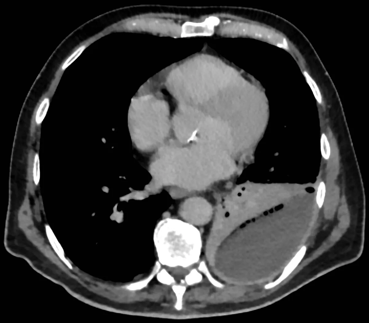 CT
CT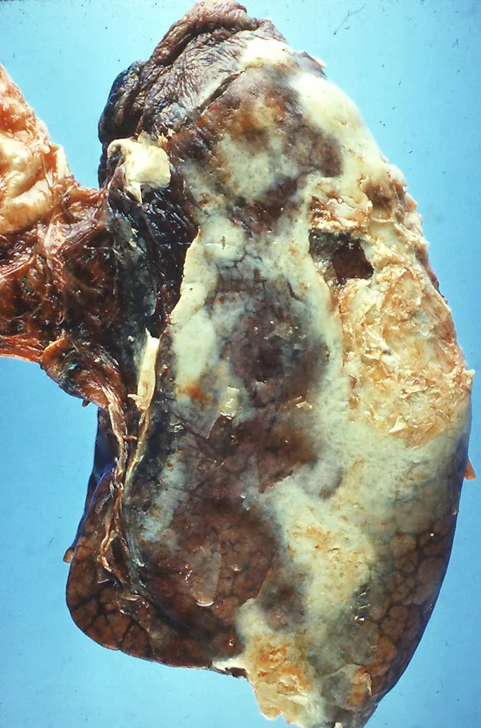 Gross specimen
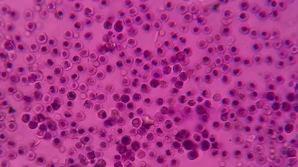 Blood smear & cytology
Definition
Pleural empyema is a collection of pus in the pleural space. It is the severe end of the spectrum of pleural infection, which is defined as bacterial entry and replication in the pleural space and includes complicated parapneumonic effusion. When an empyema spreads into the soft tissues and causes chest wall infection with external discharge, it is called empyema necessitatis.
Occurrence & epidemiology
Pleural infections are common: in the USA and the United Kingdom together more than 80,000 cases are recorded per year, with rising incidence across the Western world. About 20–57 % of patients with pneumonia have a pleural effusion at presentation, and 5–7 % of these progress to pleural infection.
Aetiopathogenesis
- Origin: usually a complication of pneumonia; however, about 30 % of pleural infections have no accompanying pneumonia on imaging. Other causes are thoracotomy, abscesses of the lung, liver or subphrenic space and penetrating injuries with secondary infection.
- Risk factors for progression of pneumonia to pleural infection: immunosuppression, diabetes, poor oral hygiene, gastresophageal reflux, alcohol excess and intravenous drug use.
- Pathogens: the spectrum differs from that of pneumonia. Staphylococcus aureus has replaced viridans streptococci as the most common pathogen, about 60 % of it MRSA. Community-acquired infections tend to be gram-positive, hospital-acquired ones gram-negative; anaerobes are important but difficult to culture. Polymicrobial infections are common, atypical organizms rare.
Clinical features
- Classic course: rather younger patients with few comorbidities, fever, rigors, acute respiratory symptoms and a non-resolving pneumonia with pleural effusion.
- Insidious course: particularly in older patients with multiple conditions, with nonspecific symptoms such as loss of weight, loss of appetite and malaise that resemble cancer and can delay the diagnosis.
- Complication: a fibrous peel around the lung (trapped lung) can restrict lung expansion.
Diagnosis
- Chest X-ray: usually the first investigation; low sensitivity for effusions, more than 10 % of significant parapneumonic effusions are missed, especially with lower lobe consolidation.
- Thoracic ultrasound: detects echogenic fluid and septations and allows safe sampling.
- Contrast-enhanced chest CT: enhancement of parietal and visceral pleura ("split pleura sign"), microbubbles, effusion volume, underlying lung changes.
- Pleural fluid sampling: the gold standard of diagnosis with assessment of appearance (purulent), biochemistry and microbiology. pH is the most useful single parameter: pH ≤ 7.2 means a high, pH > 7.2 to < 7.4 an intermediate and pH ≥ 7.4 a low likelihood of pleural infection. In the intermediate range LDH (above 900 IU/L) and glucose (≤ 4 mmol/L) help; if the pH is unreliable, a glucose below 2.2 mmol/L can be used.
- Microbiology: pleural fluid culture is positive in 30–40 %, rising to about 60 % after inoculation into blood culture bottles; blood cultures are the only positive sample in some cases.
Keep learning in the app
Further reading (open access)
Cross-references
More topics: Pulmonology
- Community-acquired pneumonia
- Tuberculosis
- Pneumothorax
- Sarcoidosis
- Lung cancer (bronchial carcinoma)
- COPD (chronic obstructive pulmonary disease)
- Pleural effusion
- Asthma
- Acute respiratory distress syndrome (ARDS)
- Legionnaires' disease
- Alpha-1 antitrypsin deficiency
- Idiopathic pulmonary fibrosis and interstitial lung disease
Note: Learning content for medical education – not a treatment recommendation and no substitute for diagnosis or treatment decisions in individual cases. Treatment and management are deliberately not covered on this page.