Pleural effusion
Board exam relevance: in 7 of 105 exam reports · rank 53- Synonyms
- fluid on the lung, water on the lung, pleural fluid, transudate, exudate, hydrothorax
- Specialty
- Internal medicine · Pulmonology
- Images
- Diagram 1 · X-ray 1 · CT 1
- Last updated
- 10/2026 · Dr. Pascal Bafteh
Contents
Images (3)
 Diagram
 X-ray
X-ray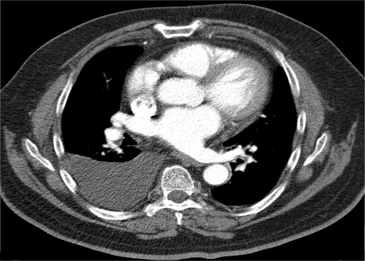 CT
CTDefinition
A pleural effusion is an increased accumulation of fluid in the pleural space. Physiologically, the pleural space contains only 10–20 mL of fluid, similar in composition to plasma but with a lower protein content.
Classification
- Transudate: caused by raised hydrostatic and/or reduced oncotic pressure.
- Exudate: caused by local processes with increased capillary permeability through which fluid, protein and cells escape.
- Special forms: empyema (pus in the pleural space), hemothorax (pleural fluid hematocrit above 50 % of blood hematocrit), chylothorax (milky, rich in triglycerides, due to damage of the thoracic duct, most commonly by lymphoma), chyliform (pseudochylous) effusion (rich in cholesterol, low in triglycerides, mainly in rheumatoid pleuritis and chronic tuberculosis).
Aetiopathogenesis
There are more than 50 causes.
- Transudates: heart failure as the most common cause (bilateral in 81 %), cirrhosis with ascites (hepatic hydrothorax, mostly right-sided), nephrotic syndrome and other forms of hypoalbuminemia, constrictive pericarditis, hypothyroidism, atelectasis.
- Exudates: most commonly infections (parapneumonic effusion, viral infections, tuberculosis), cancer (mainly lung and breast cancer and lymphoma) and pulmonary embolism (effusion in about 40 %, almost always exudative, often hemorrhagic). Other causes are acute pancreatitis, rheumatoid arthritis, systemic lupus erythematosus, drugs, esophageal rupture, subphrenic abscess, benign asbestos pleural effusion, Meigs syndrome and yellow nail syndrome.
Clinical features
- Some effusions are asymptomatic and found incidentally
- Breathlessness and/or pleuritic chest pain that is sharp on breathing; irritation of the diaphragmatic pleura can radiate to the abdomen, neck or shoulder
- Findings: diminished breath sounds, dullness to percussion and absent tactile fremitus on the side of the effusion; rapid, shallow breathing with large effusions; rarely a pleural friction rub
Diagnosis
- Thoracic ultrasound: the most sensitive method; detects small amounts of fluid, septations and pleural thickening and guides the tap.
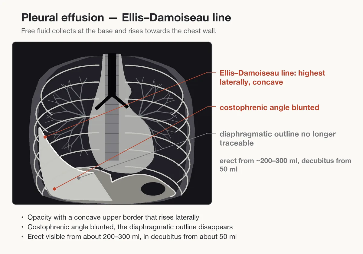
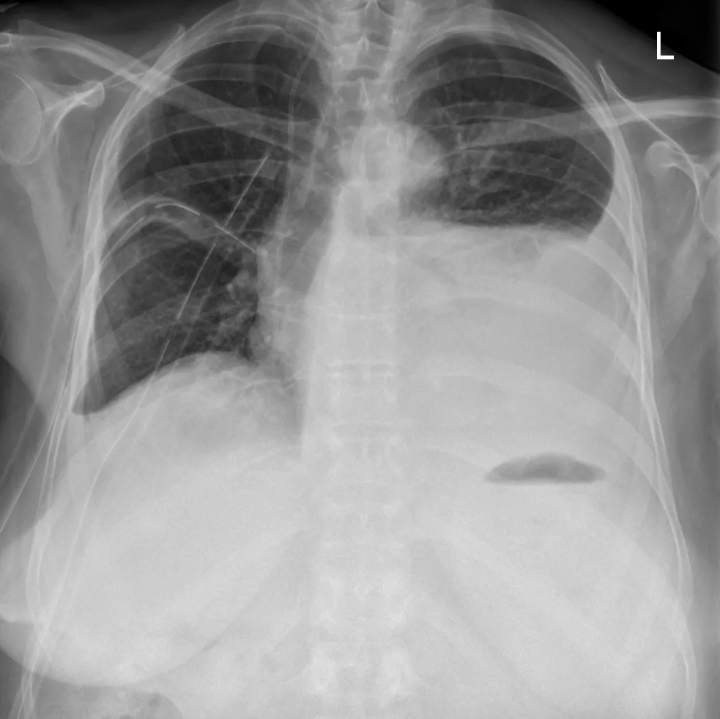
- Chest X-ray: blunting of the costophrenic angle, with large effusions opacification of the hemithorax; effusions above 4 L can opacify the whole hemithorax and shift the mediastinum to the opposite side. Loculated effusions in the fissures can mimic a tumor (pseudotumor).
- Contrast-enhanced CT: pleural enhancement, pleural nodules, hidden lung infiltrates or masses.
- Diagnostic thoracentesis: appearance (bloody, chylous, purulent, viscous), total protein, glucose, LDH, cell count with differential, Gram stain; depending on the question pH, cultures, cytology, tuberculosis markers (adenosine deaminase), amylase, triglycerides, cholesterol.
- Adenosine deaminase: values above 40 U/L have a sensitivity and specificity of 95 % for tuberculous pleuritis; raised values also occur in cancer.
Light's criteria
An exudate is present if at least one of the following criteria is met:
- Pleural fluid to serum total protein ratio ≥ 0.5
- Pleural fluid to serum LDH ratio ≥ 0.6
- Pleural fluid LDH ≥ two thirds of the upper limit of normal for serum LDH
Light's criteria identify almost all exudates but misclassify about 20 % of transudates as exudates. When a transudate is suspected (e.g. heart failure, cirrhosis), a difference between serum and pleural fluid protein of more than 3.1 g/dL (31 g/L) favors a transudate.
Keep learning in the app
Further reading (open access)
Cross-references
More topics: Pulmonology
- Community-acquired pneumonia
- Tuberculosis
- Pneumothorax
- Sarcoidosis
- Lung cancer (bronchial carcinoma)
- COPD (chronic obstructive pulmonary disease)
- Asthma
- Acute respiratory distress syndrome (ARDS)
- Legionnaires' disease
- Pleural empyema
- Alpha-1 antitrypsin deficiency
- Idiopathic pulmonary fibrosis and interstitial lung disease
Note: Learning content for medical education – not a treatment recommendation and no substitute for diagnosis or treatment decisions in individual cases. Treatment and management are deliberately not covered on this page.