CPPD disease (pseudogout, chondrocalcinosis)
- Synonyms
- pseudogout, chondrocalcinosis, calcium pyrophosphate arthritis, CPP crystal arthritis, crowned dens syndrome
- Specialty
- Internal medicine · Rheumatology & immunology
- Images
- X-ray 1 · CT 1
- Last updated
- 10/2026 · Dr. Pascal Bafteh
Contents
Images (2)
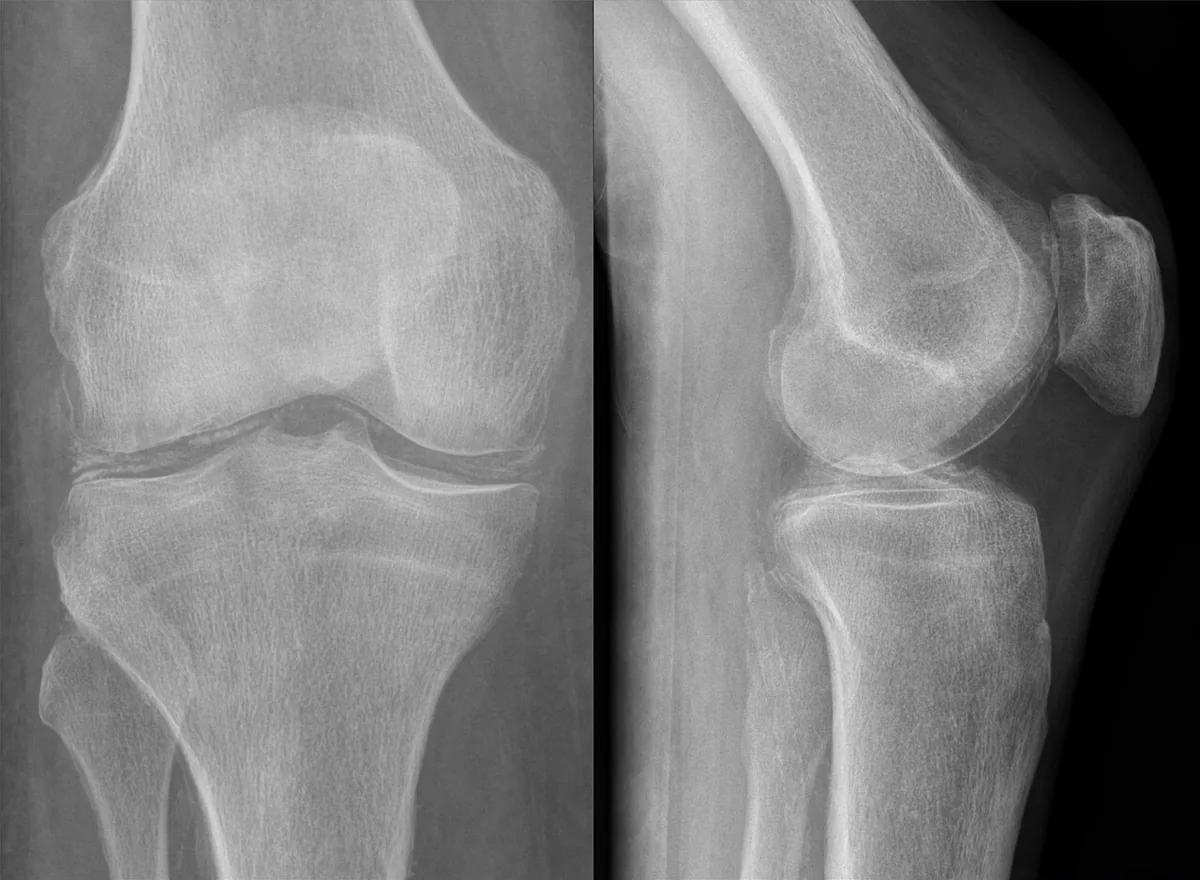 X-ray
X-ray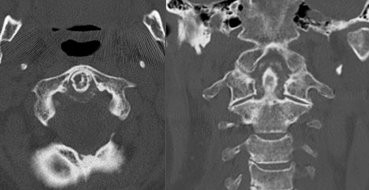 CT
CTDefinition
Calcium pyrophosphate deposition (CPPD) disease involves intra-articular and extra-articular deposition of calcium pyrophosphate crystals. Manifestations are protean: from asymptomatic calcification (chondrocalcinosis) through recurrent flares of acute arthritis (pseudogout, acute CPP crystal arthritis) to an often severe degenerative arthropathy.
Aetiopathogenesis
The cause is usually unknown; risk increases with age. Associated conditions: joint trauma (including after surgery), hypomagnesemia, hyperparathyroidism, gout, hemochromatosis, hypophosphatasia, Gitelman syndrome, X-linked hypophosphatemic rickets, and familial hypocalciuric hypercalcemia. Familial forms are usually inherited in an autosomal dominant pattern. The transport protein ANK moves pyrophosphate into the extracellular space and thereby promotes crystal formation.
Clinical features
- Acute, subacute, or chronic arthritis, usually of the knee, wrist, or other large peripheral joints; it can mimic many other forms of arthritis.
- Acute flares resemble gout but vary more in intensity and tend to be more protracted.
- Between flares, no symptoms or continuous low-grade symptoms in multiple joints, similar to rheumatoid arthritis or osteoarthritis; these patterns usually persist for life.
- Crowned dens syndrome: acute involvement at the dens of the axis with profound inflammatory neck pain and stiffness; on radiographs the dens appears "crowned" by calcification.
Diagnosis
- Arthrocentesis with polarized light microscopy: rhomboid or rod-shaped crystals, not birefringent or only weakly positively birefringent (blue parallel to the compensator axis). During a flare, the fluid is inflammatory; Gram stain and culture exclude infection, and absence of urate crystals excludes gout (both may coexist).
- Radiographs or ultrasound when synovial fluid cannot be obtained: linear or punctate calcification in articular cartilage, especially fibrocartilage (e.g., menisci); supports the diagnosis but does not exclude gout or infection. Chondrocalcinosis also occurs without flares.
- Search for underlying disorders, especially in younger patients with frequent flares: calcium, parathyroid hormone, magnesium, alkaline phosphatase, and iron studies.
Keep learning in the app
Further reading (open access)
Cross-references
More topics: Rheumatology & immunology
- Rheumatoid arthritis
- Systemic lupus erythematosus (SLE)
- Granulomatosis with polyangiitis (GPA)
- Polymyalgia rheumatica
- Gout
- Reactive arthritis
- Behçet's disease
- Giant cell arteritis (temporal arteritis)
- Septic arthritis
- Mixed connective tissue disease (Sharp syndrome)
- Psoriatic arthritis
- Uveitis in rheumatic diseases
Note: Learning content for medical education – not a treatment recommendation and no substitute for diagnosis or treatment decisions in individual cases. Treatment and management are deliberately not covered on this page.