Dermatomyositis and polymyositis
- Synonyms
- dermatomyositis, polymyositis, muscle inflammation, myositis, antisynthetase syndrome, inclusion body myositis
- Specialty
- Internal medicine · Rheumatology & immunology
- Images
- Histology 1 · Clinical 3
- Last updated
- 10/2026 · Dr. Pascal Bafteh
Contents
Images (4)
 Histology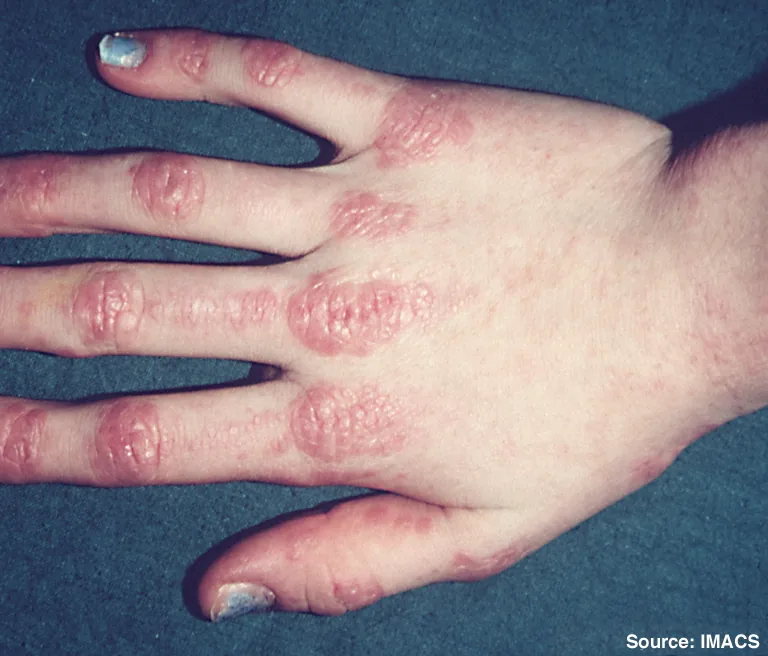
Definition
Idiopathic inflammatory myopathies are autoimmune disorders with inflammatory and degenerative changes in the muscles (e.g., polymyositis, immune-mediated necrotizing myopathy) or in the skin and muscles (dermatomyositis). The leading symptom is symmetric weakness mainly of the proximal pelvic and shoulder girdle muscles, sometimes with myalgia, atrophy, and fibrous replacement.
Classification
- Dermatomyositis (including the amyopathic form without muscle involvement): with typical skin changes
- Antisynthetase syndrome: myositis, interstitial lung disease, nonerosive arthritis, fever, "mechanic's hands", Raynaud syndrome
- Immune-mediated necrotizing myopathy: e.g., anti-SRP- and anti-HMGCR-associated (including statin-associated); severe weakness, very high CK, dysphagia
- Inclusion body myositis: older age, slowly progressive, also distal muscles (hands, feet)
- Polymyositis: myositis without skin involvement, now diagnosed less often
- Overlap myositis: together with systemic lupus erythematosus or systemic sclerosis
Occurrence & epidemiology
Except for inclusion body myositis, most forms are more common in females. The disorders occur most commonly between ages 40 and 60 or, in children, between ages 5 and 15.
Aetiopathogenesis
An autoimmune reaction against muscle tissue in genetically susceptible people is suspected; familial clustering and HLA associations (e.g., haplotype HLA-DRB1*03-DQA1*05-DQB1*02) are known. Possible triggers are viral infections, certain drugs, and cancer; in dermatomyositis and necrotizing myopathy, a tumor may incite myositis via antigens shared by tumor and muscle.
Dermatomyositis is considered a complement-mediated vasculopathy with immune complex deposition, polymyositis a direct T cell-mediated muscle injury; in necrotizing myopathy, macrophages and myophagocytosis predominate.
Clinical features
- Muscle weakness over weeks to months: difficulty raising the arms above the head, climbing stairs, and rising from a seated position; weakness of the neck flexors; hands, feet, eye, and facial muscles are spared except in inclusion body myositis.
- Dysphagia and risk of aspiration due to involvement of pharyngeal and upper esophageal muscles; in severe cases dysphonia and diaphragmatic weakness.
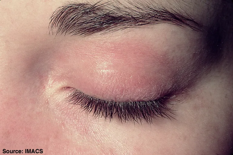
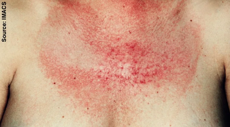
- Skin in dermatomyositis: violaceous periorbital edema (heliotrope rash), Gottron papules over the metacarpophalangeal and proximal interphalangeal joints, V sign and shawl sign, nail-fold changes, calcinosis (especially the juvenile form).
- Lungs: interstitial lung disease, especially with antisynthetase antibodies; cardiac involvement with pericarditis or cardiomyopathy.
- Polyarthralgias, Raynaud syndrome, fever, fatigue, weight loss.
- Cancer association, mainly in dermatomyositis (e.g., ovary, cervix, lung, bladder, prostate; in Asia nasopharynx).
Histology
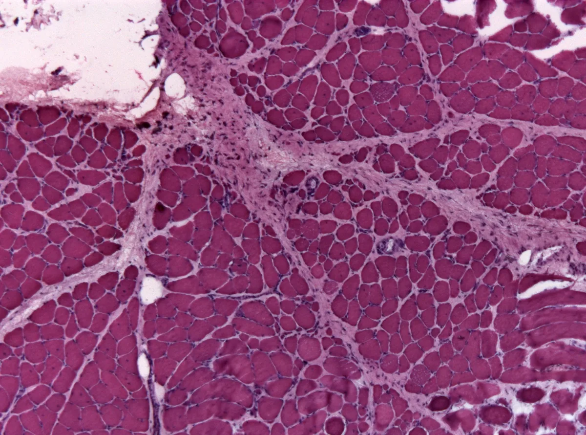
Muscle biopsy shows chronic inflammation with fiber degeneration and regeneration. Polymyositis shows predominantly endomysial CD8-positive T-cell infiltrates without vasculopathy, dermatomyositis perifascicular, B cell-predominant infiltrates with perivascular inflammation.
Diagnosis
- Muscle enzymes: creatine kinase (CK, most specific), aldolase, aminotransferases; troponin I elevated with cardiac involvement.
- Autoantibodies: ANA in up to 80 %; myositis-specific antibodies assign subtypes: anti-Jo-1 and other antisynthetase antibodies (myositis, interstitial lung disease), anti-Mi-2 (severe skin disease), anti-MDA5 (amyopathic dermatomyositis, severe interstitial lung disease, skin ulcers), anti-TIF1γ and anti-NXP-2 (increased cancer risk), anti-SRP and anti-HMGCR (necrotizing myopathy), anti-cN1A (inclusion body myositis), anti-PM-Scl and anti-Ku (overlap).
- Electromyography and MRI of affected muscles: exclude neurogenic causes and show muscle edema as a suitable biopsy site.
- Lungs and heart: radiographs, CT, pulmonary function tests, and echocardiography when interstitial lung disease or pulmonary hypertension is suspected; swallowing study with dysphagia.
- Search for cancer, mainly in dermatomyositis.
Keep learning in the app
Further reading (open access)
Cross-references
More topics: Rheumatology & immunology
- Rheumatoid arthritis
- Systemic lupus erythematosus (SLE)
- Granulomatosis with polyangiitis (GPA)
- Polymyalgia rheumatica
- Gout
- Reactive arthritis
- Behçet's disease
- Giant cell arteritis (temporal arteritis)
- Septic arthritis
- Mixed connective tissue disease (Sharp syndrome)
- Psoriatic arthritis
- Uveitis in rheumatic diseases
Note: Learning content for medical education – not a treatment recommendation and no substitute for diagnosis or treatment decisions in individual cases. Treatment and management are deliberately not covered on this page.