Axial spondyloarthritis (ankylosing spondylitis)
- Synonyms
- ankylosing spondylitis, Bechterew disease, axSpA, inflammatory back pain, bamboo spine
- Specialty
- Internal medicine · Rheumatology & immunology
- Images
- X-ray 2 · MRI 2
- Last updated
- 10/2026 · Dr. Pascal Bafteh
Contents
Images (4)
 X-ray
X-ray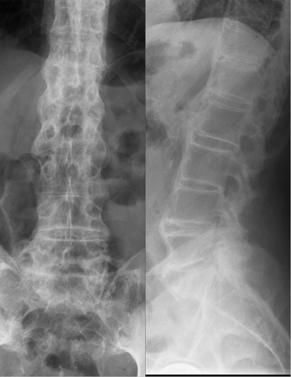 X-ray
X-ray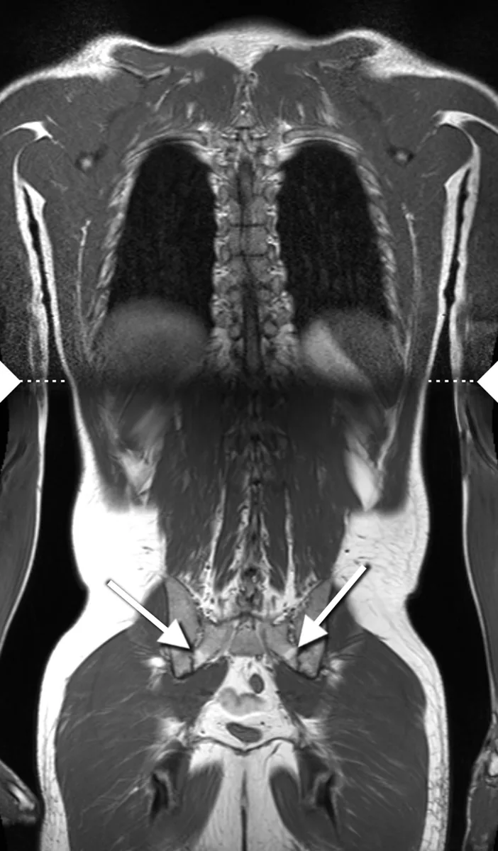 MRI
MRI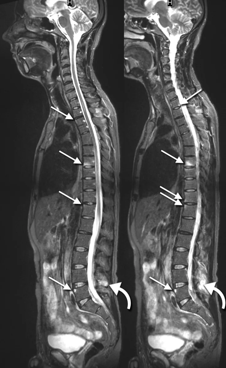 MRI
MRIDefinition
Axial spondyloarthritis is a chronic inflammatory disease mainly of the axial skeleton (sacroiliac joints and spine). Its prototype is ankylosing spondylitis, a systemic disorder with nocturnal back pain, morning stiffness, increasing kyphosis, involvement of large peripheral joints and digits, constitutional symptoms, and possible aortitis, cardiac conduction abnormalities, and anterior uveitis.
Classification
- Radiographic axial spondyloarthritis (ankylosing spondylitis): sacroiliitis on radiographs, typically bilateral
- Nonradiographic axial spondyloarthritis: clinically similar, without typical sacroiliitis on radiographs
- Peripheral form: predominantly peripheral joint involvement
ASAS classification criteria (back pain for at least 3 months, onset before age 45):
- Imaging arm: sacroiliitis on radiographs or MRI plus at least 1 SpA feature
- Clinical arm: HLA-B27 plus at least 2 other SpA features
- SpA features: inflammatory back pain, arthritis, heel enthesitis, uveitis, dactylitis, psoriasis, inflammatory bowel disease, good response to NSAIDs, family history, HLA-B27, elevated CRP
Occurrence & epidemiology
Men are affected about three times more often than women. The disease usually begins between ages 20 and 40. It is 10 to 20 times more common among first-degree relatives than in the general population.
Aetiopathogenesis
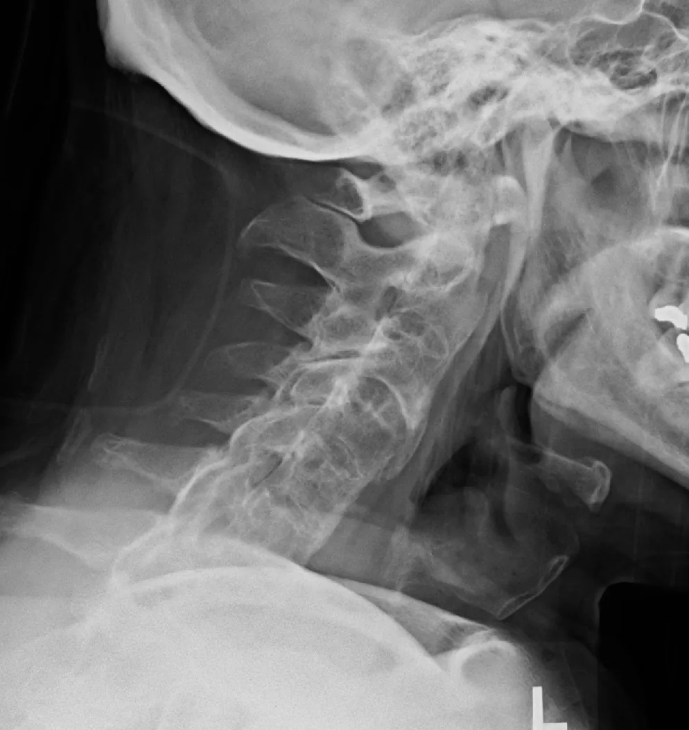
A strong genetic component is shown by the association with HLA-B27: the allele is present in about 90 % of White patients but, depending on ethnicity, also in up to 10 % of the general population. Inflammation affects the sacroiliac joints, spine, and tendon insertions; over time, subchondral erosions, sclerosis, syndesmophytes, ligamentous calcification, and finally bony fusion (ankylosis) develop.
Clinical features
- Inflammatory back pain: the most common symptom, often nocturnal; onset by age 40, gradual, morning stiffness, improvement with activity, duration of at least 3 months.
- Reduced chest expansion due to costovertebral involvement; occasionally fatigue, weight loss, anemia.
- Increasing kyphosis, loss of lumbar lordosis, in late stages fixed bent-forward posture.
- Arthritis of hips and shoulders, dactylitis, Achilles and patellar tendinitis; onset in peripheral joints especially in children and women.
- Systemic manifestations in about 25 %: recurrent acute anterior uveitis, aortic insufficiency, aortitis, pericarditis, conduction abnormalities, rarely apical pulmonary fibrosis.
- Nonspecific colitis in about 30 %; neurologic complications from vertebral fractures or cauda equina syndrome.
Diagnosis
- Laboratory tests: ESR and CRP inconsistently elevated; rheumatoid factor and antinuclear antibodies negative; HLA-B27 supports the diagnosis but has low predictive value.
- Radiographs of the sacroiliac joints and lumbar spine: initially pseudo-widening due to subchondral erosions, then sclerosis, joint space narrowing, and fusion, symmetric. In the spine, vertebral squaring with corner sclerosis ("shiny corner"), syndesmophytes, in late stages bamboo spine.
- Pelvic MRI: shows early inflammation (bone marrow edema) and structural changes before radiographs become abnormal.
- Typical constellation: young man with nocturnal back pain, prolonged morning stiffness, reduced chest expansion, enthesitis, or unexplained anterior uveitis.
Keep learning in the app
Further reading (open access)
Cross-references
More topics: Rheumatology & immunology
- Rheumatoid arthritis
- Systemic lupus erythematosus (SLE)
- Granulomatosis with polyangiitis (GPA)
- Polymyalgia rheumatica
- Gout
- Reactive arthritis
- Behçet's disease
- Giant cell arteritis (temporal arteritis)
- Septic arthritis
- Mixed connective tissue disease (Sharp syndrome)
- Psoriatic arthritis
- Uveitis in rheumatic diseases
Note: Learning content for medical education – not a treatment recommendation and no substitute for diagnosis or treatment decisions in individual cases. Treatment and management are deliberately not covered on this page.