Bartholin cyst and abscess
- Synonyms
- Bartholin cyst, Bartholin abscess
- Specialty
- Gynaecology · Vulva & vagina
- Images
- Clinical 4
- In the app
- 1 flashcards · GynFuchs
- Last updated
- 10/2026 · Dr. Pascal Bafteh
Contents
Images (4)


Definition
Bartholin's Cyst and Abscess
- Location: The Bartholin's gland duct opens in the posterior third of the introitus, at approximately the 5 and 7 o'clock positions.
- Pathogenesis of the cyst: It forms due to obstruction of the excretory duct with secretion build-up.
- Clinical presentation of the abscess: A characteristic finding is a tense, extremely painful, reddened swelling that impedes walking.
- A Bartholin cyst forms when the duct of the Bartholin gland is obstructed and mucus accumulates; infection of a cyst or of the gland itself produces a Bartholin abscess.
- The two pea-sized (about 0.5 cm) glands, normally not palpable, lie at the lower posterior sides of the vaginal opening, at about 4 and 8 o'clock; their 2.0–2.5 cm ducts open between the labia minora and the hymenal edge.
Occurrence & epidemiology
Epidemiology
- Cysts and abscesses usually occur after the onset of puberty; Bartholin cysts develop in about 3 % of women, usually in their 20s.
- Abscesses are almost three times more common than cysts alone.
- Incidence decreases after the menopause, when the possibility of malignant transformation becomes more relevant, especially in recurrent or complex cases.
Aetiopathogenesis
Aetiology and risk factors
- Bartholin abscesses are polymicrobial; Escherichia coli is the single most common pathogen identified on culture.
- Rarely, the infection is caused by a sexually transmitted pathogen such as Neisseria gonorrhoeae.
Clinical features
- Most cysts are asymptomatic, non-tender, unilateral and palpable near the vaginal orifice; large cysts can cause pressure or pain and interfere with sexual activity, sitting or walking.
- Abscesses cause severe vulvar pain and sometimes fever; they are tender and typically erythematous, and surrounding cellulitis and vaginal discharge may also occur.
Diagnosis
Typical imaging findings
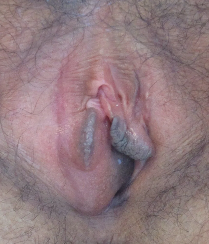
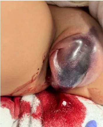
- Clinical finding: A smooth, tense, unilateral swelling bulges from the posterior third of the labium majus and displaces the introitus sideways. The overlying skin is not red and the other side is normal.
- The diagnosis is usually made by examination of the vulva; typical findings are a unilateral, medially protruding mass in the posterior vulva with vulvar asymmetry and, in abscesses, overlying erythema, induration or fluctuance.
- Bartholin gland carcinoma is rare; a solid or fixed mass raises suspicion of malignancy.
Keep learning in the app
Further reading (selection)
Cross-references
Note: Learning content from the GynFuchs app (flashcards, exam questions, image cases) for medical education – not a treatment recommendation and no substitute for diagnosis or treatment decisions in individual cases. Treatment and management are deliberately not covered on this page.