Anogenital warts (condylomata acuminata)
Exam relevance: in 4 of 197 board exam reports · rank 111
- Synonyms
- genital warts, anogenital warts
- Specialty
- Gynaecology · Vulva & vagina
- Images
- Ultrasound 2 · Colposcopy 3 · Clinical 2 · Histology & cytology 1
- Exam relevance
- 4 of 197 reports · rank 111
- In the app
- 2 flashcards · GynFuchs
- Last updated
- 10/2026 · Dr. Pascal Bafteh
Contents
Images (8)
 Ultrasound
 Colposcopy
 Histology & cytology
 Ultrasound
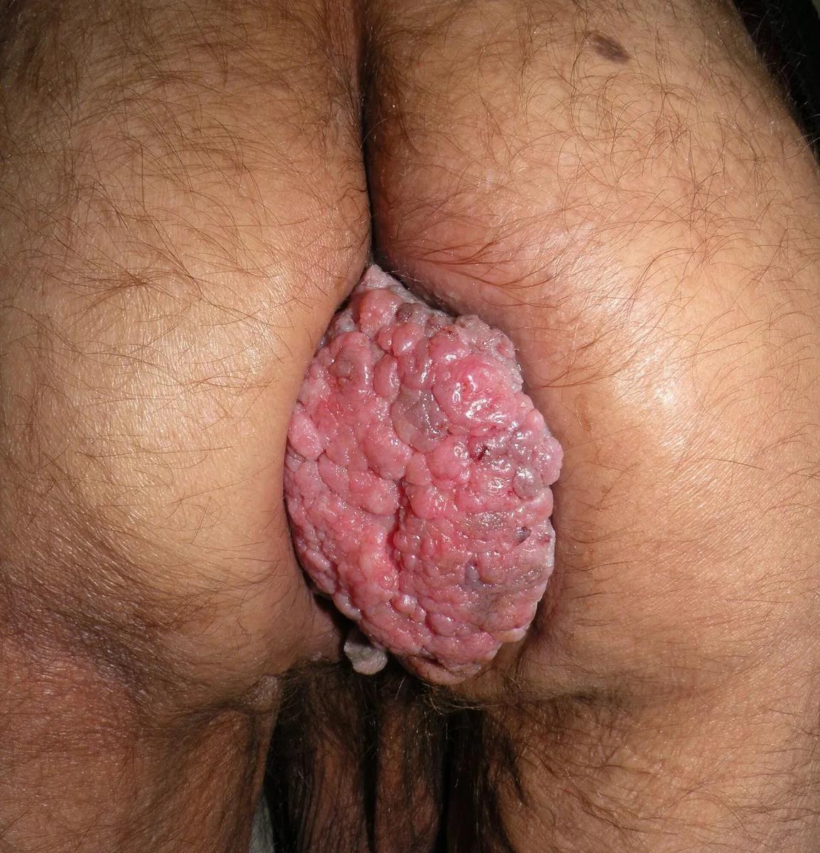
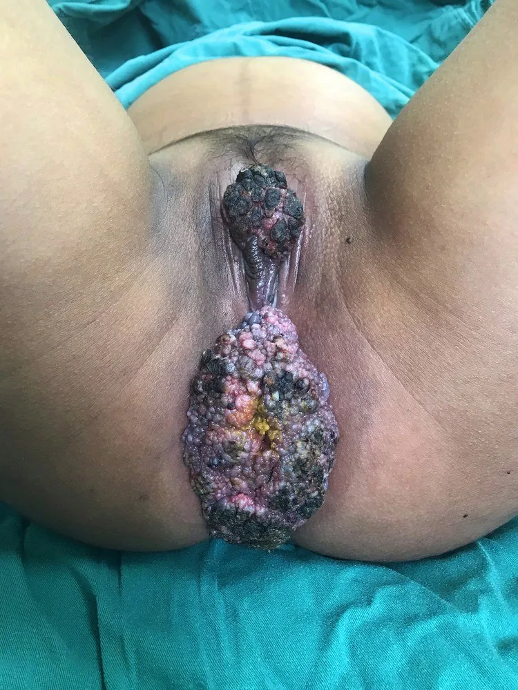 Colposcopy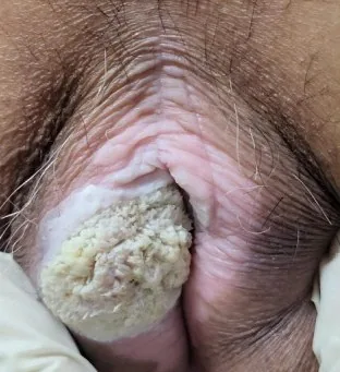
 Colposcopy
Definition
- Anogenital warts (condylomata acuminata) are the clinical manifestation of a sexually transmitted infection with certain HPV types.
Occurrence & epidemiology
Epidemiology
- HPV is the most common sexually transmitted infection; most HPV infections clear spontaneously within 1–2 years.
- About 90 % of those who contract HPV do not develop genital warts.
Aetiopathogenesis
Aetiology and risk factors
- HPV types 6 and 11 cause about 90 % of genital warts; condylomas are benign, and the HPV types that cause cervical or anal cancer are different ones.
- Transmission occurs through direct skin-to-skin contact with an infected person, usually during sex.
- With reduced cell-mediated immunity, for example in pregnancy or HIV infection, genital warts may persist and spread widely.
Clinical features
Condylomata on the cervix
- Causative agents: HPV 6 and 11.
- At colposcopy they appear acetowhite with pointed, finger-like projections.
Condylomata on the vulva
- Pathogen: Condylomata acuminata are caused by low-risk HPV types, mainly 6 and 11.
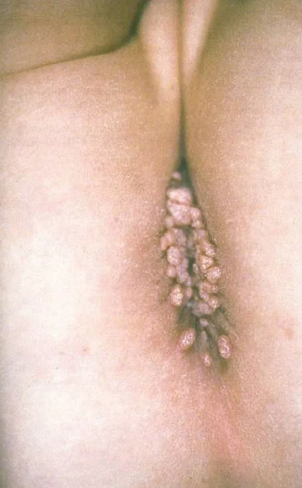
- Warts appear after an incubation period of 1–6 months; in women they occur most commonly on the vulva, vaginal wall, cervix and perineum, and the urethra and anal region may also be affected.
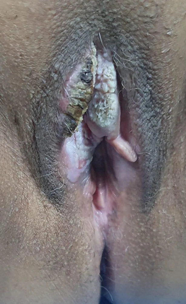
- Lesions are papular, verrucous or sessile, flat or exophytic, and may coalesce into cauliflower-like plaques; they range from millimetre-sized papules to centimetre-sized masses and may bleed with friction.
- They are usually asymptomatic; some patients have itching, burning or discomfort.
Histology
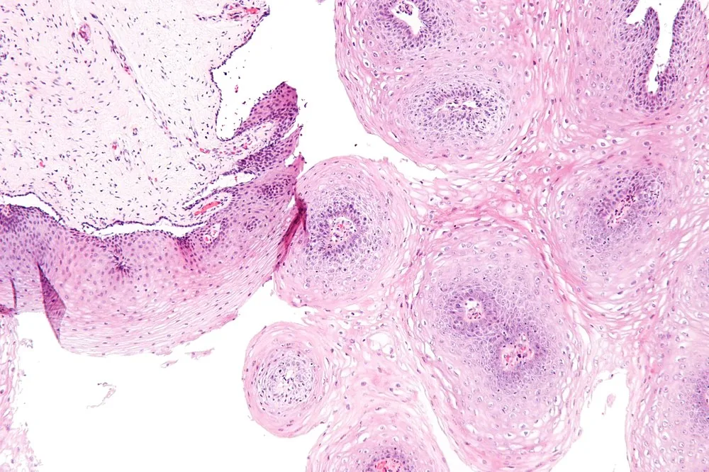
- Histologically, HPV-related lesions show papillomatosis and koilocytosis.
Diagnosis
Typical imaging findings
- Colposcopy: A whitish, finely papillary, cauliflower-like growth with a lobulated surface sits on the cervix. It is soft, not ulcerated and does not bleed spontaneously; the surrounding epithelium is normal.
- Clinical finding: Numerous small, pointed, soft skin-coloured papules stand in beds perineally and perianally; some are cauliflower-like and lobulated. The surroundings are quiet, with no ulceration or induration.
- Clinical finding: A firm, verrucous exophytic growth with a fissured, partly crusted surface sits on the labium majus. It is clearly larger and firmer than ordinary condylomas and grows in depth; the surrounding tissue is displaced.
- Clinical finding: A palm-sized, cauliflower-like growth made of densely packed reddish fronds surrounds the anus. It grows on a broad base and pushes the surrounding skin apart; the surface is macerated but not ulcerated.
- The diagnosis is usually made by visual inspection.
- In atypical, bleeding, ulcerated or persistent warts, a biopsy serves to exclude carcinoma.
- After application of 3–5 % acetic acid, lesions turn white, which makes even small lesions easier to detect at colposcopy.
Keep learning in the app
Further reading (selection)
Cross-references
Note: Learning content from the GynFuchs app (flashcards, exam questions, image cases) for medical education – not a treatment recommendation and no substitute for diagnosis or treatment decisions in individual cases. Treatment and management are deliberately not covered on this page.