Velamentous cord insertion and umbilical cord anomalies
Exam relevance: in 2 of 197 board exam reports · rank 144
- Specialty
- Obstetrics · Pregnancy disorders
- Images
- Endoscopy & gross 5
- Exam relevance
- 2 of 197 reports · rank 144
- In the app
- 1 flashcards · GynFuchs
- Last updated
- 10/2026 · Dr. Pascal Bafteh
Contents
Images (5)
 Endoscopy & gross
 Endoscopy & gross
 Endoscopy & gross
 Endoscopy & gross
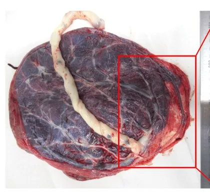 Endoscopy & gross
Definition
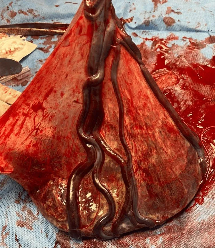
- In velamentous cord insertion the cord inserts into the membranes rather than the placenta; the umbilical vessels run for some distance between the membranes to the placenta.
- This segment of the vessels lacks the protection of Wharton's jelly, which normally surrounds the umbilical vessels, and is therefore susceptible to rupture and thrombosis.
Classification
Insertions
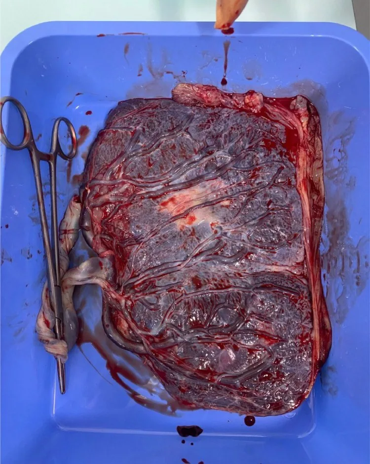
- Normal is a central or paracentral cord insertion into the placenta.
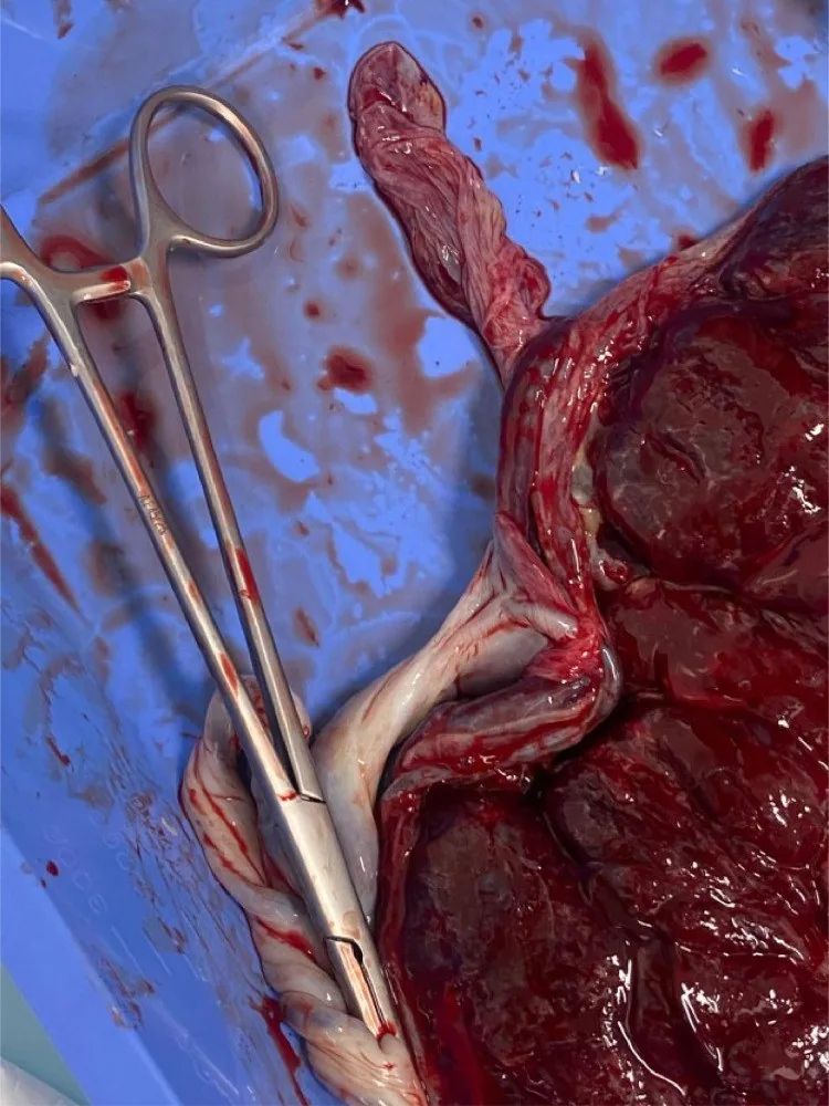
- Marginal insertion means insertion at the placental edge ("battledore").
- Velamentous insertion means the vessels run without Wharton's jelly across the membranes to the placenta and is the main precursor of vasa praevia.
- Insertion anomalies are commoner in multiple pregnancy and after assisted reproduction.
Further variants
- A succenturiate placenta is an accessory lobe joined to the main placenta by vessels; its danger lies in retention of the accessory lobe with secondary bleeding, and in vasa praevia.
- A single umbilical artery is associated with malformations and growth restriction and calls for a careful anomaly scan.
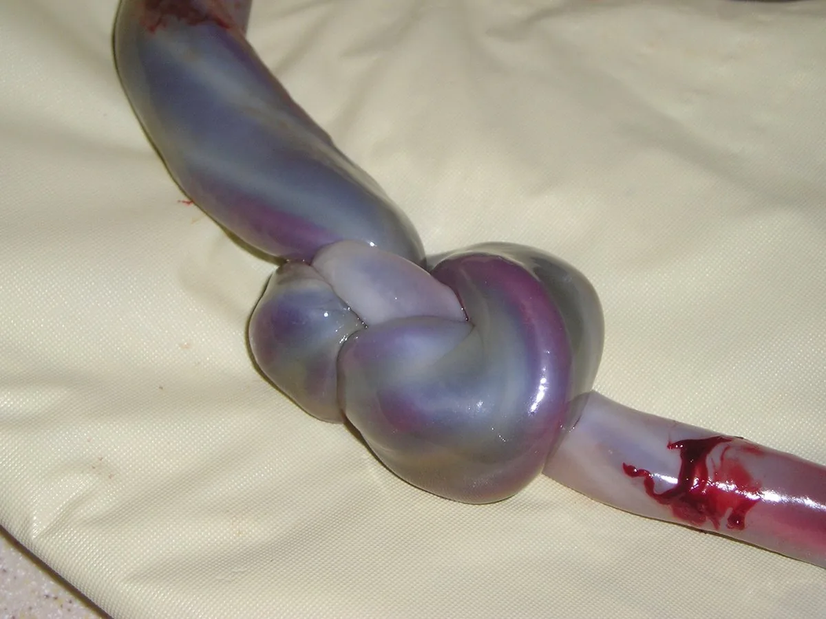
- A true knot is a genuine knot in the cord with a raised but overall low risk of strangulation; a false knot, by contrast, is a harmless loop of vessels or a heap of Wharton's jelly.
- The Grannum classification describes the sonographic placental grades 0 to III.
- Other insertion variants are marginal cord insertion, in which the cord inserts at the edge of the placenta, and the rare furcate insertion, in which the vessels separate from the cord before inserting into the placenta.
Occurrence & epidemiology
Epidemiology
- Velamentous insertion is reported in about 1% of pregnancies; in studies its frequency ranges from 0.4% to 11% in singletons and from 1.6% to 40% in twins.
- Marginal insertion occurs in about 7% of pregnancies.
Clinical features
Clinical features and complications
- Velamentous insertion is closely associated with vasa praevia, in which unprotected vessels run close to the internal os and may rupture, which can lead to fatal fetal exsanguination.
- It is associated with fetal growth restriction, congenital anomalies and retained placenta; rupture or thrombosis of the vessels can lead to preterm birth or intrauterine fetal death.
- In singletons it is associated mainly with preterm birth and emergency caesarean section, in twins with perinatal mortality, although the increase in risk is usually only low to moderate.
Diagnosis
- The placental cord insertion is visualised at routine ultrasound, particularly in twins; insertion abnormalities are best diagnosed with Doppler ultrasound.
Keep learning in the app
Further reading (selection)
- StatPearls: Embryology, Umbilical Cord (NCBI Bookshelf)
- Med Sci Monit 2019: Modern Ultrasonography of the Umbilical Cord (PMC, open access)
- Syst Rev 2020: Velamentous cord insertion – rapid review of incidence, risk factors, adverse outcomes and screening (PMC, open access)
- DocCheck Flexikon, Insertio velamentosa
Cross-references
Note: Learning content from the GynFuchs app (flashcards, exam questions, image cases) for medical education – not a treatment recommendation and no substitute for diagnosis or treatment decisions in individual cases. Treatment and management are deliberately not covered on this page.