Multiple pregnancy
Exam relevance: in 25 of 197 board exam reports · rank 30
- Synonyms
- twin pregnancy, twins
- Specialty
- Obstetrics · Pregnancy disorders
- Images
- Ultrasound 3
- Exam relevance
- 25 of 197 reports · rank 30
- In the app
- 3 flashcards · GynFuchs
- Last updated
- 10/2026 · Dr. Pascal Bafteh
Contents
Images (3)
 Ultrasound
 Ultrasound
 Ultrasound
Definition
- A multiple pregnancy is the simultaneous intrauterine development of two or more fetuses; it carries higher risks than a singleton pregnancy.
Classification
Timing of division is decisive
- A very early division in the first three days leads to dichorionic-diamniotic (DCDA) twins.
- The most common form in monozygotic twins is the monochorionic-diamniotic (MCDA) configuration, which occurs with division between the fourth and eighth day.
- A late division between the eighth and 13th day results in monochorionic-monoamniotic (MCMA) twins, while an even later division leads to conjoined twins.
Why the foetal membranes are so important
- The clinically decisive factor is not zygosity, but chorionicity, as a shared placenta implies vascular anastomoses and thus specific risks like TTTS.
- Specific risks of monochorionic pregnancies are twin-to-twin transfusion syndrome (TTTS), TAPS, selective foetal growth restriction, and the TRAP sequence.
- The detection of different sexes reliably rules out monochorionicity.
- Monoamniotic twins additionally have the high risk of umbilical cord entanglement and knotting.
- Dizygotic twins arise from two fertilised oocytes and are always dichorionic; monozygotic twins arise from division of a single fertilised oocyte.
- In monozygotic twins the timing of division determines placentation: within the first 72 hours dichorionic-diamniotic, between days 4 and 8 monochorionic-diamniotic, after day 8 monochorionic-monoamniotic.
- About 75% of monozygotic pregnancies are monochorionic, and about 2% of these are monoamniotic.
Occurrence & epidemiology
Epidemiology
- Twins account for 2–4% of all births; in the United States in 2021 there were 21.3 twin births per 1000 live births.
- The frequency of monozygotic twins is largely constant at 3.5–4 per 1000 births; regional differences are mainly due to dizygotic twins.
- The twin rate has risen because of higher maternal age and assisted reproduction.
Aetiopathogenesis
Two paths to twins
- Dizygotic twins arise from two eggs and two sperm, with risk factors such as maternal age, family history, and assisted reproduction increasing the frequency and account for the majority of twin pregnancies.
- In terms of the foetal membranes, they are always dichorionic and diamniotic (DCDA).
- Monozygotic twins arise from the division of a single zygote; their rate is approximately constant worldwide.
Aetiology and risk factors
- Risk factors for dizygotic twins include geographical origin, Black ethnicity, multiparity, advanced maternal age, low socioeconomic status, family history and assisted reproduction.
Clinical features
Maternal risks
- The most common maternal risks, in addition to hyperemesis, anaemia and gestational diabetes, primarily include hypertensive disorders of pregnancy and pre-eclampsia.
- Due to the overstretched uterus, there is a significantly increased risk of postpartum haemorrhage postpartum.
- Further risks include a higher caesarean section rate, thromboembolism and a high psychosocial burden.
Fetal risks
- The main problem for the fetuses is preterm birth, which affects the majority of twins; in addition, growth restrictions and discordances are more common.
- Malformations, umbilical cord complications and intrauterine fetal death lead to significantly increased perinatal mortality.
More facts from the study questions
- Dizygotic twins arise from two fertilised oocytes.
- They always develop with separate placentas and amniotic sacs (dichorionic-diamniotic).
- Monochorionic twins arise from a single zygote and are therefore genetically identical.
- Genetically identical twins are always of the same sex.
- Dizygotic twins constitute the majority of all twin pregnancies and are always dichorionic-diamniotic (DCDA).
- While the monochorionic-diamniotic (MCDA) type is the most common form in monozygotic twins, it is not the most common overall.
- The timing of a monozygotic zygote's division determines the configuration of the membranes.
- A very early division (day 1-3) results in a dichorionic-diamniotic configuration, which is sonographically indistinguishable from dizygotic twins.
- Monoamniotic twins share a single amniotic sac, allowing their umbilical cords to move freely.
- This carries a high risk of entanglement and knot formation, which can compromise fetal perfusion.
Clinical features and complications
- Maternal problems include more frequent hyperemesis, dilutional anaemia, hypertensive disorders (up to two- to 3.5-fold), gestational diabetes, preterm labour, rupture of membranes, placental abruption and postpartum atony and haemorrhage.
- Perinatal mortality is two to three times higher than in singletons, mainly due to preterm birth, growth restriction, low birth weight and intrapartum hypoxia; malformations and chromosomal disorders affect about 2% of twin pregnancies.
- Specific to monochorionic twins are twin-to-twin transfusion syndrome and selective growth restriction, which affects 10–15% of monochorionic pregnancies.
- Monochorionic monoamniotic twins are rare (about 2% of monochorionic pregnancies); umbilical cord accidents are typical, occur in 48–80% and are associated with high perinatal mortality.
Diagnosis
Chorionicity on ultrasound
- The lambda or twin-peak sign, a triangular projection of chorionic tissue at the base of the dividing membrane, is indicative of a dichorionic pregnancy.
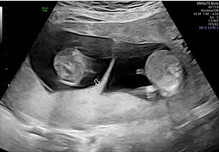
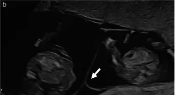
- The T-sign, where the thin membrane inserts perpendicularly into the placenta, is, in contrast, an indication of a monochorionic diamniotic pregnancy.
Typical imaging findings
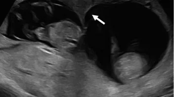
- Ultrasound: A dividing membrane runs between the two sacs. At its insertion on the placenta a wedge of tissue extends into the membrane, producing a triangular lambda shape; the membrane appears thick.
- Chorionicity is determined by ultrasound early, within the first 13 weeks; the lambda sign is typical of dichorionic pregnancies.
- Selective growth restriction is diagnosed when the weight difference exceeds 25% and one fetus is below the 10th centile.
Keep learning in the app
Further reading (selection)
Cross-references
Note: Learning content from the GynFuchs app (flashcards, exam questions, image cases) for medical education – not a treatment recommendation and no substitute for diagnosis or treatment decisions in individual cases. Treatment and management are deliberately not covered on this page.