Intrauterine fetal death (stillbirth)
Exam relevance: in 9 of 197 board exam reports · rank 75
- Synonyms
- stillbirth, fetal death
- Specialty
- Obstetrics · Pregnancy disorders
- Images
- Ultrasound 1
- Exam relevance
- 9 of 197 reports · rank 75
- Last updated
- 10/2026 · Dr. Pascal Bafteh
Contents
Images (1)
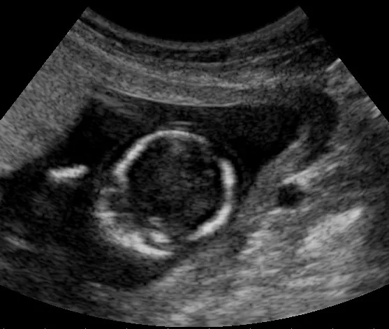 Ultrasound
Definition
- Intrauterine fetal death is the death of the fetus before complete birth, shown by the fetus not breathing or showing any other sign of life such as heartbeat or cord pulsation afterwards; the boundary with miscarriage is defined differently between countries.
- In the United States, stillbirth is fetal death from 20 weeks or from 350 g; the WHO definition uses 28 weeks.
Occurrence & epidemiology
Epidemiology
- Worldwide there are almost 2 million stillbirths each year, about 98% of them in low- and middle-income countries; fewer than 5% are recorded.
- Worldwide, no cause is reported for 76% of stillbirths.
Aetiopathogenesis
Aetiology and risk factors
- Common causes include intrapartum complications, hypertension, diabetes, infection, congenital and genetic anomalies, placental dysfunction and pregnancy continuing beyond 40 weeks.
- Maternal risk factors include pre-eclampsia, trauma, acquired thrombophilias, poorly controlled diabetes, thyroid disease, morbid obesity, substance misuse, smoking, sepsis and age under 20 or 35 years and over; a previous stillbirth increases the risk.
- Placental and fetal causes include placental abruption, chorioamnionitis, fetomaternal haemorrhage, twin-to-twin transfusion syndrome, cord accidents, vasa praevia, chromosomal abnormalities, major malformations, non-immune hydrops and multiple pregnancy.
Clinical features
Clinical features and complications
- If a fetus that has died in late pregnancy remains in the uterus for weeks, consumptive coagulopathy or even disseminated intravascular coagulation may develop.
Diagnosis
- The diagnosis is clinical; on ultrasound, previously documented cardiac activity is absent.
- Investigation of the cause includes external examination of the baby, autopsy, karyotyping and microarray analysis, placental examination, maternal full blood count, Kleihauer-Betke test, antiphospholipid antibodies, TSH, HbA1c, TORCH serology, syphilis screening and toxicology screening; often the cause nevertheless remains unclear.
Keep learning in the app
Further reading (selection)
Cross-references
Note: Learning content from the GynFuchs app (flashcards, exam questions, image cases) for medical education – not a treatment recommendation and no substitute for diagnosis or treatment decisions in individual cases. Treatment and management are deliberately not covered on this page.