Monochorionic twin pregnancy
Exam relevance: in 9 of 197 board exam reports · rank 75
- Specialty
- Obstetrics · Pregnancy disorders
- Images
- Ultrasound 1
- Exam relevance
- 9 of 197 reports · rank 75
- In the app
- 1 flashcards · GynFuchs
- Last updated
- 10/2026 · Dr. Pascal Bafteh
Contents
Images (1)
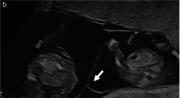 Ultrasound
Definition
- A monochorionic twin pregnancy is a twin pregnancy with a single shared placenta; up to one third of all twin pregnancies are monochorionic.
Classification
- Monochorionic diamniotic twins (one placenta, two amniotic sacs) are distinguished from monochorionic monoamniotic twins (one placenta, one amniotic sac).
Aetiopathogenesis
Aetiology and pathogenesis
- Monochorionic twins are monozygotic; diamniotic twins arise from splitting between days 3 and 8, monoamniotic twins between days 8 and 13 after fertilisation.
- In nearly all monochorionic placentas, vascular anastomoses connect the two fetal circulations; their angioarchitecture determines the risk profile.
Clinical features
Specifics of monochorionicity
- Specific risks in monochorionic pregnancies are twin-to-twin transfusion syndrome (TTTS), TAPS, selective fetal growth restriction and the TRAP sequence.
- In monoamniotic pregnancies, an additional serious risk is umbilical cord entanglement.
- If one fetus dies in a monochorionic pregnancy, an acute transfusion into the deceased fetus occurs via the anastomoses, which can lead to hypotension with death or neurological damage in the surviving twin.
- Approximately one in every four to five monochorionic twin pregnancies develops twin-to-twin transfusion syndrome.
Clinical features and complications
- Complications unique to monochorionic pregnancy are twin-to-twin transfusion syndrome (TTTS), twin anaemia-polycythaemia sequence (TAPS), TRAP sequence, monoamnionicity and conjoined twins.
- After intrauterine death of one twin in a monochorionic pregnancy, the co-twin also dies in about 15%, and postnatal cranial imaging is abnormal in about 34%.
Diagnosis
- On ultrasound, the T sign indicates monochorionicity and the lambda sign dichorionicity; chorionicity is best determined in the first trimester.
- According to Quintero, TTTS is present with a deepest vertical pocket of at most 2 cm in the donor and at least 8 cm in the recipient.
- TAPS is recognised prenatally by discordant middle cerebral artery Doppler findings, for example a peak systolic velocity of at least 1.5 MoM in the donor and at most 0.8 MoM in the recipient.
Keep learning in the app
Further reading (selection)
Cross-references
Note: Learning content from the GynFuchs app (flashcards, exam questions, image cases) for medical education – not a treatment recommendation and no substitute for diagnosis or treatment decisions in individual cases. Treatment and management are deliberately not covered on this page.