Uterine malformations (septate, bicornuate uterus)
Exam relevance: in 3 of 197 board exam reports · rank 125
- Specialty
- Gynaecology · Uterus & endometrium
- Images
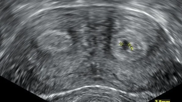
- Ultrasound 1
- Exam relevance
- 3 of 197 reports · rank 125
- Last updated
- 10/2026 · Dr. Pascal Bafteh
Contents
Images (1)
Definition
- Congenital uterine anomalies result from abnormal formation, canalisation or fusion of the Müllerian ducts or from defective resorption of the midline septum.
- A bicornuate uterus results from incomplete fusion of the Müllerian ducts, typically between weeks 6 and 10, and has two separate endometrial cavities with a single cervix.
Classification
- The ASRM classification (most recently 2021, nine categories) and the 2013 ESHRE/ESGE classification are in use; neither is universally accepted.
- For a septate uterus ASRM 2021 requires an indentation deeper than 1 cm with an angle under 90° and a normal external shape, ESHRE/ESGE an indentation-to-wall-thickness ratio over 50%.
Occurrence & epidemiology
Epidemiology
- Congenital uterine anomalies are found in about 5.5% of unselected women, 8% of infertile women and 13.3% of women with miscarriage.
- The septate uterus accounts for about 35% of anomalies, with a prevalence of 0.2–2.3% in women of reproductive age.
Clinical features
- With a septate uterus, infertility, recurrent miscarriage, preterm birth and fetal malpresentation are more frequent.
- A bicornuate uterus is likewise associated with infertility, recurrent pregnancy loss, preterm labour and malpresentation.
Diagnosis
- Hysteroscopy is the gold standard for the septate uterus; 3D transvaginal ultrasound shows the cavity and the external fundal contour in the coronal plane.
- 3D ultrasound or MRI is used to distinguish a bicornuate from a septate uterus.
- In a meta-analysis the pooled sensitivity and specificity of 2D transvaginal ultrasound for septate uterus were 83% and 99%.
Keep learning in the app
Further reading (selection)
Cross-references
Note: Learning content from the GynFuchs app (flashcards, exam questions, image cases) for medical education – not a treatment recommendation and no substitute for diagnosis or treatment decisions in individual cases. Treatment and management are deliberately not covered on this page.