Endometrial cancer
Exam relevance: in 35 of 197 board exam reports · rank 18
- Synonyms
- uterine cancer, womb cancer
- Specialty
- Gynaecology · Uterus & endometrium
- Images
- Endoscopy & gross 3 · Ultrasound 4
- Exam relevance
- 35 of 197 reports · rank 18
- In the app
- 4 flashcards · GynFuchs
- Last updated
- 10/2026 · Dr. Pascal Bafteh
Contents
Images (7)
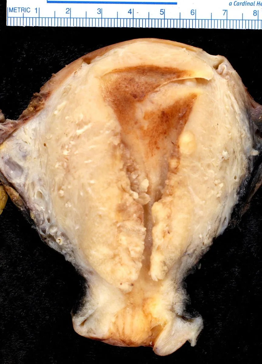 Endoscopy & gross
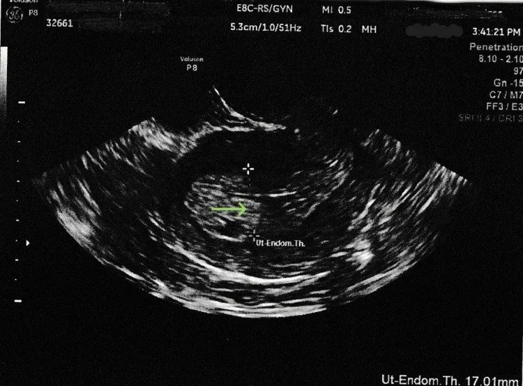 Ultrasound
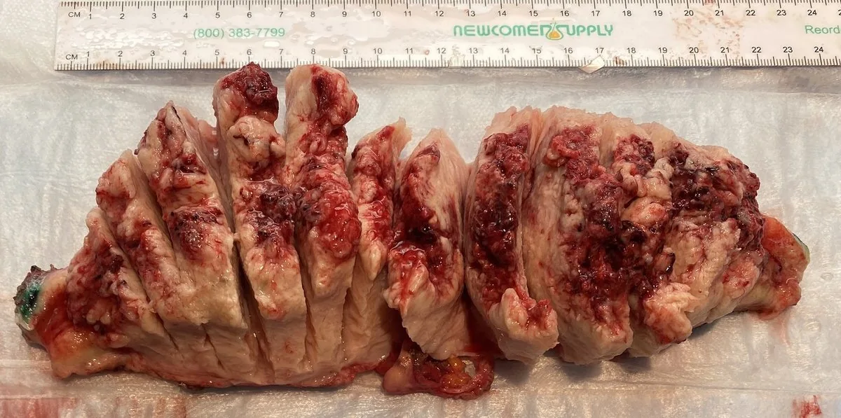 Endoscopy & gross
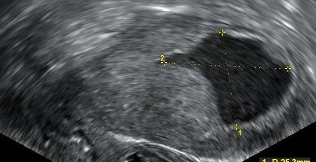 Ultrasound
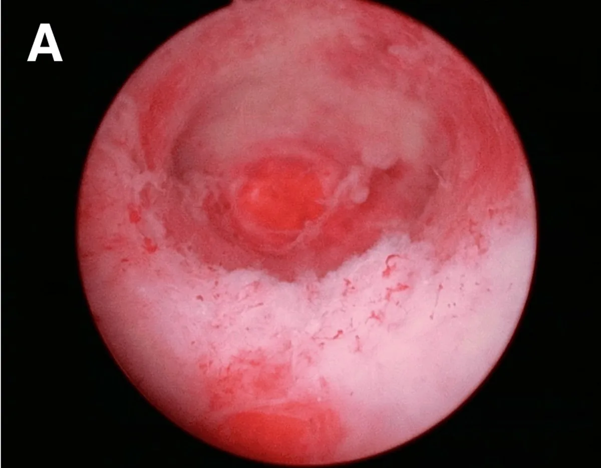 Endoscopy & gross
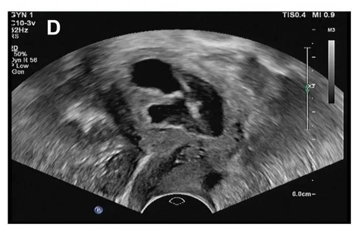 Ultrasound
 Ultrasound
Classification
Stages
- FIGO I: Stage FIGO I means tumour confinement to the corpus uteri.
- FIGO II: Stage FIGO II means invasion of the cervical stroma.
- FIGO III: Stage FIGO III means local/regional spread.
- FIGO IIIC: Stage FIGO IIIC1 denotes pelvic, IIIC2 para-aortic LN metastases.
- FIGO IV: Stage FIGO IV means invasion of bladder/bowel mucosa; distant metastases.
- New feature: A new feature in the current FIGO classification is integration of molecular groups/LVSI.
Why it was introduced
- The old classification into Type I and Type II was prognostic, but not predictive.
- The molecular classification is now part of the staging classification.
- For the classification, the following are determined: IHC for MMR/p53, molecular for POLE.
Four groups
- POLE-mutated means POLE exonuclease domain mutation.
- MMRd means loss of mismatch repair proteins.
- p53abn means aberrant p53 staining, high copy number.
- NSMP stands for “no specific molecular profile”.
- L1CAM is prognostic marker for NSMP group (poorer).
Aetiopathogenesis
Basic Principle
- Pathogenesis: The majority of endometrial carcinomas develop with oestrogen dominance without progestogen.
- Oestrogen-independent carcinomas: In contrast, a portion of carcinomas, especially the serous types, develop oestrogen-independent with atrophy.
Risk Factors
- Metabolic factors: Metabolic risk factors include obesity, type 2 diabetes mellitus, hypertension.
- Reproductive factors: Reproductive risk factors are nulliparity, early menarche, late menopause.
- Hereditary factors: A hereditary risk factor is Lynch syndrome (mismatch repair defect).
Clinical features
Basic Principle
- Principle: Any bleeding after menopause is considered requiring workup until cancer is excluded.
- Most common finding: The most common finding is endometrial atrophy.
- Carcinoma incidence: A carcinoma is found in approx. 10% of cases, rising with age.
- Definition: Menopause is defined as final period (after 12 months of amenorrhea).
More facts from the study questions
- Lynch syndrome is a hereditary risk factor.
- It is characterised by a defect in the mismatch repair genes.
- An investigation, e.g. by hysteroscopy, is only performed if bleeding occurs.
- The grading of endometrioid carcinoma is based on the proportion of solid, non-glandular tumour areas.
- G1 and G2 are grouped as low-grade, G3 as high-grade.
- Aggressive, non-endometrioid types (serous, clear cell, etc.) are classified as high-grade (G3) by definition.
- Endometrioid adenocarcinoma is by far the most common histological subtype of endometrial carcinoma.
- Infiltration of the cervical stroma, with the tumour confined to the uterus, defines FIGO stage II.
- FIGO stage I is confined to the uterine corpus.
- Molecular classification is no longer just an additional prognostic factor.
Histology
Histological Types
- Most common type: The most common histological type of endometrial carcinoma is endometrioid adenocarcinoma.
- Aggressive types: The aggressive, non-endometrioid types include serous, clear cell, undifferentiated ca, carcinosarcoma.
- Serous carcinoma: Serous carcinoma is p53-aberrant with atrophy.
- Carcinosarcoma: Carcinosarcoma is now considered carcinoma with sarcomatoid differentiation.
Grading
- Grading criterion: The grading of endometrioid carcinoma is based on the proportion of solid, non-glandular tumour areas.
- Grade classification: G1 and G2 are classified together as low grade, G3 as high grade.
- Non-endometrioid types: The non-endometrioid types are considered high grade, regardless of the growth pattern.
Diagnosis
Staging Investigations
- Pelvic MRI: The pelvic MRI is primarily used for assessing myometrial invasion/cervical involvement.
Examination
- Clinical examination: The examination procedure includes inspection, speculum, palpation, vaginal ultrasound.
- Cervical work-up: Cytological and colposcopic investigation is performed if bleeding is from cervical canal or smear is abnormal.
Endometrial Thickness
- Cut-off value with bleeding: The cut-off value for postmenopausal bleeding is an endometrium of 3–4 mm.
- Fluid in the cavity: Postmenopausal fluid in the uterine cavity is a finding requiring workup (suspected carcinoma).
Typical imaging findings
- Ultrasound: The cavity is filled by a chaotically structured mass, partly hypoechoic and partly echogenic. Over a long stretch the border between endometrium and myometrium can no longer be identified; the uterine wall looks irregular there.
- Ultrasound: The cavity is filled by a 17 mm inhomogeneous mass with irregular margins. The interface with the myometrium can no longer be traced in places; internal echoes are mixed hypo- and hyperechoic.
Keep learning in the app
Further reading (selection)
Cross-references
Note: Learning content from the GynFuchs app (flashcards, exam questions, image cases) for medical education – not a treatment recommendation and no substitute for diagnosis or treatment decisions in individual cases. Treatment and management are deliberately not covered on this page.