Endometrial hyperplasia
Exam relevance: in 10 of 197 board exam reports · rank 66
- Specialty
- Gynaecology · Uterus & endometrium
- Images
- Ultrasound 2 · Endoscopy & gross 1
- Exam relevance
- 10 of 197 reports · rank 66
- In the app
- 1 flashcards · GynFuchs
- Last updated
- 10/2026 · Dr. Pascal Bafteh
Contents
Images (3)
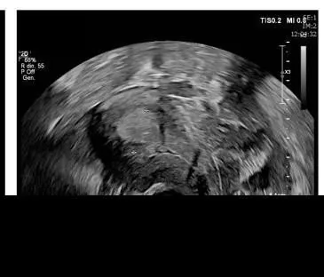 Ultrasound
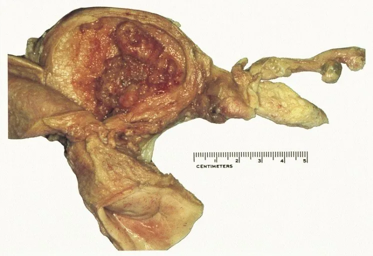 Endoscopy & gross
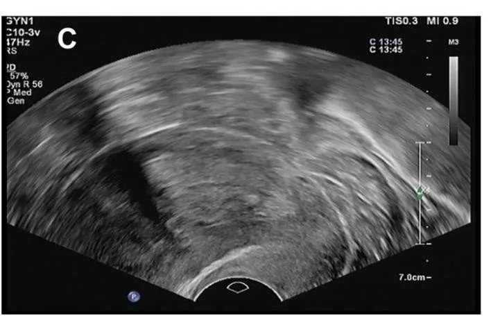 Ultrasound
Definition
- Endometrial hyperplasia is an abnormal proliferation of endometrial glands with an increased gland-to-stroma ratio compared with proliferative endometrium.
Classification
- WHO classification: The WHO distinguishes between hyperplasia without atypia & atypical hyperplasia/EIN.
- Cause: The cause is unopposed estrogen effect.
- Progression risk without atypia: The risk of progression for hyperplasia without atypia is at < 5% over 20 years.
- The 2014 WHO classification, retained in 2020, distinguishes two categories: hyperplasia without atypia and atypical hyperplasia or endometrial intraepithelial neoplasia (EIN), both terms being synonymous.
- The atypical form is the precursor of endometrioid adenocarcinoma; EIN is regarded as a mutationally activated, monoclonal pre-malignancy.
Aetiopathogenesis
Aetiology and risk factors
- The usual cause is oestrogen stimulation without adequate progesterone opposition.
- Risk factors are early menarche, late menopause, nulliparity, obesity, diabetes mellitus, PCOS, functional tumours such as granulosa cell tumours, and Lynch syndrome.
Clinical features
- Most women present with abnormal uterine bleeding; hyperplasia has been estimated to account for about 15% of postmenopausal bleeding.
- About 40% of women with EIN are diagnosed with endometrial carcinoma within 12 months, most likely because of a coexisting carcinoma not sampled at biopsy.
Histology
- EIN criteria are: gland area exceeding stromal area, cytology differing from the background, lesion diameter over 1 mm, and exclusion of mimics and carcinoma.
Diagnosis
Typical imaging findings
- Ultrasound: The endometrium is thickened along its whole length and heterogeneous, without a demarcable focal lesion. The border with the myometrium remains smooth and continuous; no invasion of the uterine wall.
- Ultrasound: The endometrium is uniformly thickened along the whole cavity and measures more than 20 mm between the calipers. It stays echogenic and homogeneous; the border to the myometrium is smooth and preserved throughout.
- Ultrasound may show a thickened endometrium; the diagnosis is made histologically.
- A biopsy may under-sample the lesion; hyperplasia may also be shed with menstruation or regress spontaneously.
Keep learning in the app
Further reading (selection)
- New concepts for an old problem: the diagnosis of endometrial hyperplasia (Hum Reprod Update 2017, PubMed Central)
- StatPearls: Endometrial Hyperplasia (NCBI Bookshelf)
- Atypical Endometrial Hyperplasia and Concurrent Cancer: A Comprehensive Overview on a Challenging Clinical Condition (Cancers (Basel) 2024, PubMed Central)
- DocCheck Flexikon, Endometriumhyperplasie
Cross-references
Note: Learning content from the GynFuchs app (flashcards, exam questions, image cases) for medical education – not a treatment recommendation and no substitute for diagnosis or treatment decisions in individual cases. Treatment and management are deliberately not covered on this page.