Endometrial polyp
Exam relevance: in 1 of 197 board exam reports · rank 171
- Specialty
- Gynaecology · Uterus & endometrium
- Images
- Ultrasound 3 · Endoscopy & gross 2 · Histology & cytology 1
- Exam relevance
- 1 of 197 reports · rank 171
- In the app
- 1 flashcards · GynFuchs
- Last updated
- 10/2026 · Dr. Pascal Bafteh
Contents
Images (6)
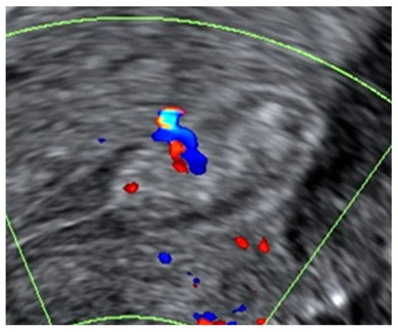 Ultrasound
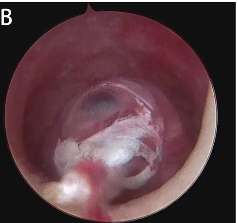 Endoscopy & gross
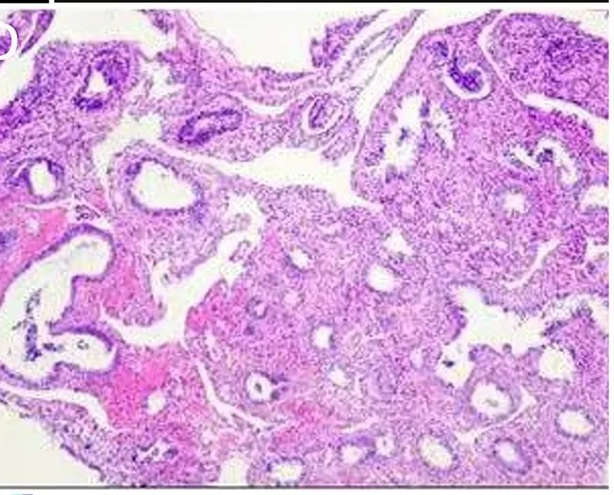 Histology & cytology
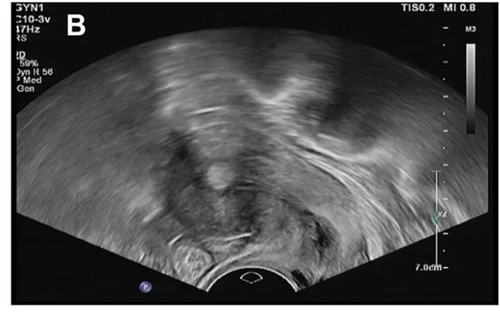 Ultrasound
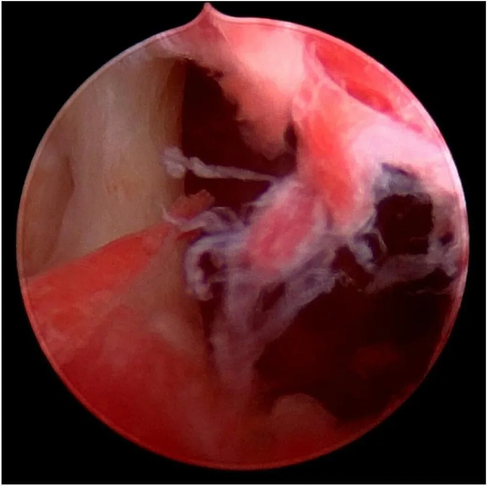 Endoscopy & gross
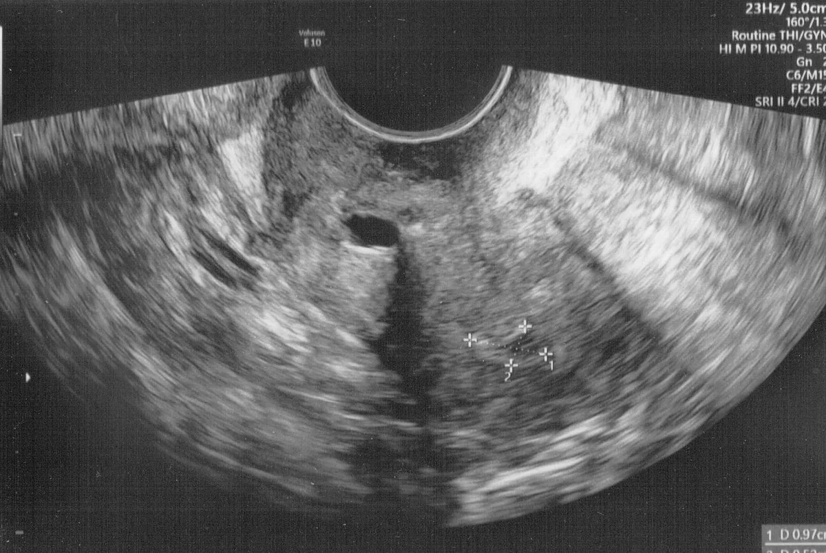 Ultrasound
Definition
- Endometrial polyps are localised proliferations of endometrial glands, stroma and a central vascular core projecting into the uterine cavity, ranging from a few millimetres to several centimetres.
- Pedunculated polyps are attached by a narrow stalk, sessile polyps by a broad base without a stalk.
Occurrence & epidemiology
Epidemiology and pathogenesis
- Polyps are among the most frequent pathological findings in the uterus; incidence peaks between 40 and 49 years.
- Their development is linked to oestrogen stimulation.
Clinical features
- Many polyps are asymptomatic; in one study 82% of histologically confirmed polyps caused no symptoms.
- Polyps are a common cause of abnormal uterine bleeding and have been associated with infertility.
- Only a very small fraction, about 1%, shows hyperplastic or malignant change.
Diagnosis
Imaging of the endometrial polyp
- On ultrasound, it is recognised by a circumscribed, echogenic structure, smooth border.
- On Doppler, a typical finding is a single feeding vessel.
- It can be better visualised by hydrosonography.
Typical imaging findings
- Ultrasound: A circumscribed echogenic mass with small cystic inclusions lies within the cavity. It stands out sharply from the surrounding endometrium; the myometrium beneath is smooth and intact.
- Ultrasound: A rounded, echogenic structure inside the cavity is distinguishable from the surrounding endometrium and has been measured with calipers. The cavity line is widened at this point; the surrounding myometrium is homogeneous.
- Transvaginal ultrasound is the primary initial tool, showing polyps as hyperechogenic lesions with a regular contour.
- Colour Doppler may show the typical single feeding vessel; intrauterine contrast at sonohysterography may outline small polyps missed on greyscale ultrasound.
- Polyps may coexist with diffuse endometrial pathology such as hyperplasia or carcinoma; a tissue diagnosis requires histology.
Keep learning in the app
Further reading (selection)
Cross-references
Note: Learning content from the GynFuchs app (flashcards, exam questions, image cases) for medical education – not a treatment recommendation and no substitute for diagnosis or treatment decisions in individual cases. Treatment and management are deliberately not covered on this page.