Postmenopausal bleeding
Exam relevance: in 18 of 197 board exam reports · rank 38
- Synonyms
- PMB
- Specialty
- Gynaecology · Uterus & endometrium
- Images
- Ultrasound 3
- Exam relevance
- 18 of 197 reports · rank 38
- In the app
- 2 flashcards · GynFuchs
- Last updated
- 10/2026 · Dr. Pascal Bafteh
Contents
Images (3)
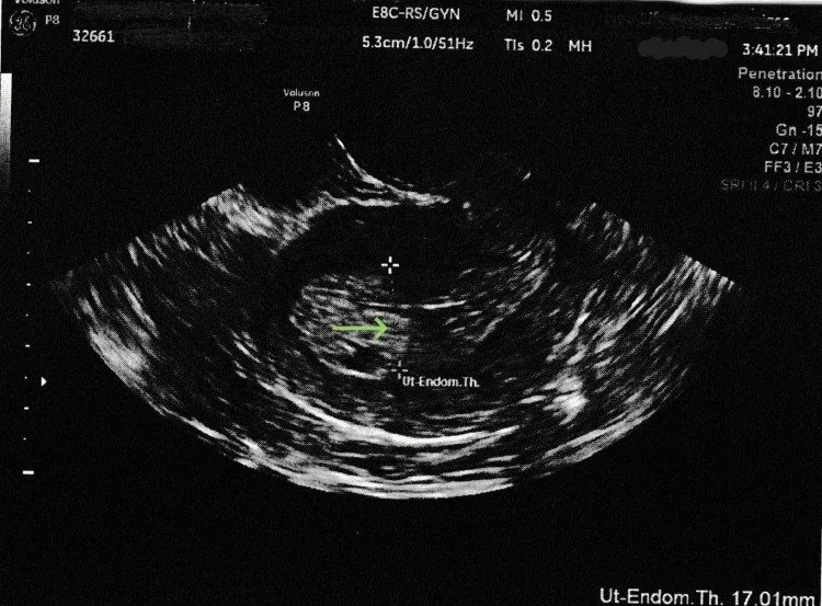 Ultrasound
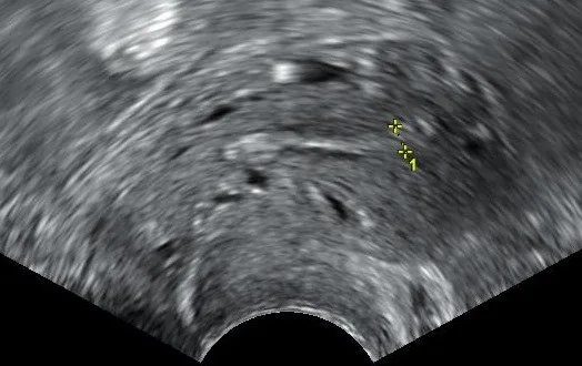 Ultrasound
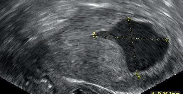 Ultrasound
Definition
Basic Principle
- Principle: Any bleeding after menopause is considered requiring workup until cancer is excluded.
- Most common finding: The most common finding is endometrial atrophy.
- Carcinoma incidence: A carcinoma is found in approx. 10% of cases, rising with age.
- Definition: Menopause is defined as final period (after 12 months of amenorrhea).
- Postmenopausal bleeding is abnormal uterine bleeding occurring more than one year after the permanent cessation of menstruation due to loss of ovarian follicular activity.
- The menopause is diagnosed clinically after one year of amenorrhoea; bleeding after this point is considered abnormal.
- In the United States the average age at menopause is 51 years.
Occurrence & epidemiology
Epidemiology
- Postmenopausal bleeding is the reason for about two-thirds of all gynaecological office visits by postmenopausal women.
- About 10% of women with postmenopausal bleeding have an intrauterine malignancy.
- Conversely, around 90% of postmenopausal women with endometrial carcinoma had vaginal bleeding.
Aetiopathogenesis
Causes and risk factors
- The commonest cause is atrophy of the endometrium or lower genital tract; other common causes are endometrial hyperplasia and polyps.
- Risk factors for type I endometrial carcinoma are obesity, older age, early menarche, late menopause, hypertension, diabetes and Lynch syndrome.
- Type I cancers are endometrioid and account for 70–80% of endometrial carcinomas; type II cancers are non-endometrioid and less often associated with these risk factors.
Clinical features
Atrophy as a source of bleeding
- What is seen on speculum examination: pale, smooth, vulnerable mucosa.
- Typical accompanying symptoms: dryness, burning, itching, dyspareunia.
- A wet mount shows: parabasal cells, elevated pH.
- Why the atrophy develops: oestrogen deficiency: thinner epithelium, glycogen loss.
- Additional consequences: urinary tract infections and urgency.
More facts from the study questions
- Atrophy of the endometrium and vagina is the most common cause of postmenopausal bleeding.
- In recurrent postmenopausal bleeding, an invasive investigation is mandatory.
- This applies even if the endometrial thickness measured by ultrasound is unremarkable.
- Fluid in the uterine cavity can indicate cervical stenosis with an underlying carcinoma.
- This finding therefore always requires investigation postmenopausally.
- Endometrial carcinoma is found in approximately one in ten cases of postmenopausal bleeding.
- The proportion of carcinomas increases with the patient's age.
- In a patient with postmenopausal bleeding, invasive investigation is performed from an endometrial thickness of 3 to 4 mm.
- Without bleeding, the cut-off is about 5 mm.
- Oestrogen deficiency leads to thinning of the epithelium, revealing deeper cell layers (parabasal cells).
Diagnosis
Examination
- Clinical examination: The examination procedure includes inspection, speculum, palpation, vaginal ultrasound.
- Cervical work-up: Cytological and colposcopic investigation is performed if bleeding is from cervical canal or smear is abnormal.
Endometrial Thickness
- Cut-off value with bleeding: The cut-off value for postmenopausal bleeding is an endometrium of 3–4 mm.
- Fluid in the cavity: Postmenopausal fluid in the uterine cavity is a finding requiring workup (suspected carcinoma).
Typical imaging findings
- Ultrasound: The endometrial stripe is thin, smoothly bordered and homogeneous throughout; the measurement is well below 5 mm. No fluid collection, no focal mass, and the border with the myometrium is intact everywhere.
- Transvaginal ultrasound is an efficient, non-invasive method for detecting endometrial pathology.
- With a pre-test probability of 10%, an endometrium below 4 mm lowers the probability of carcinoma to about 2.4%, below 5 mm to about 5%.
- Histological confirmation is obtained by endometrial biopsy or D&C.
Keep learning in the app
Further reading (selection)
- A Clinicopathological Evaluation of Postmenopausal Bleeding and Its Correlation with Risk Factors for Developing Endometrial Hyperplasia and Cancer: A Hospital-Based Prospective Study (J Midlife Health 2019, PubMed Central)
- StatPearls: Postmenopausal Bleeding (NCBI Bookshelf)
- DocCheck Flexikon, Endometriumkarzinom
- DocCheck Flexikon, Kolpitis
Cross-references
Note: Learning content from the GynFuchs app (flashcards, exam questions, image cases) for medical education – not a treatment recommendation and no substitute for diagnosis or treatment decisions in individual cases. Treatment and management are deliberately not covered on this page.