Uterine fibroids (leiomyoma)
Exam relevance: in 42 of 197 board exam reports · rank 9
- Synonyms
- fibroid, fibroids, uterine leiomyoma, myoma
- Specialty
- Gynaecology · Uterus & endometrium
- Images
- Endoscopy & gross 4 · Ultrasound 6
- Exam relevance
- 42 of 197 reports · rank 9
- In the app
- 4 flashcards · GynFuchs
- Last updated
- 10/2026 · Dr. Pascal Bafteh
Contents
Images (10)
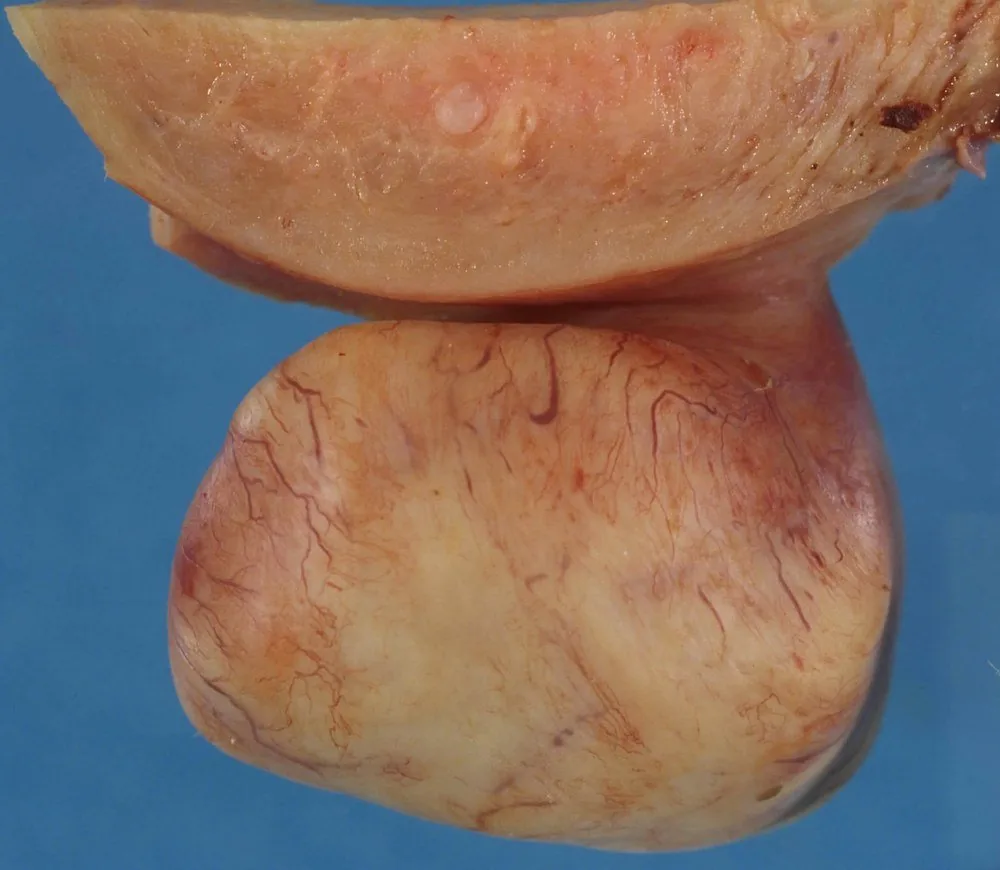 Endoscopy & gross
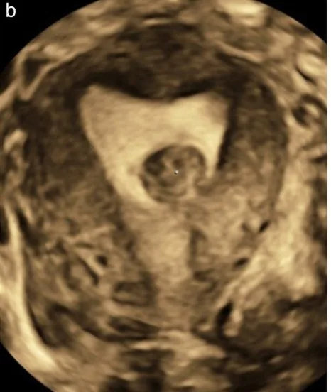 Ultrasound
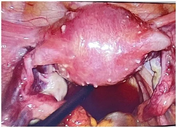 Endoscopy & gross
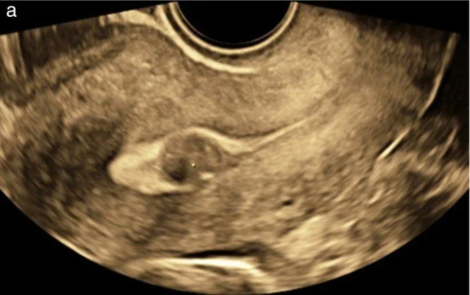 Ultrasound
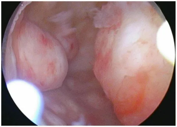 Endoscopy & gross
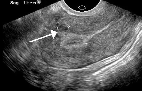 Ultrasound
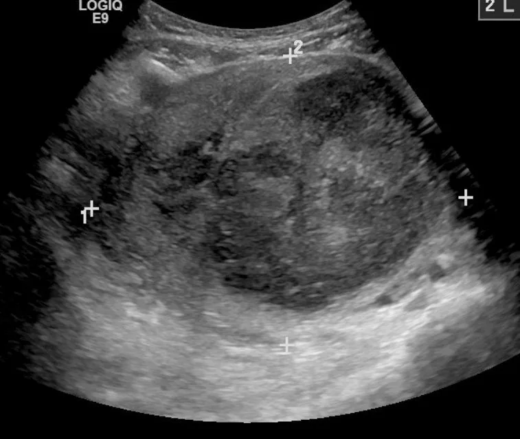 Ultrasound
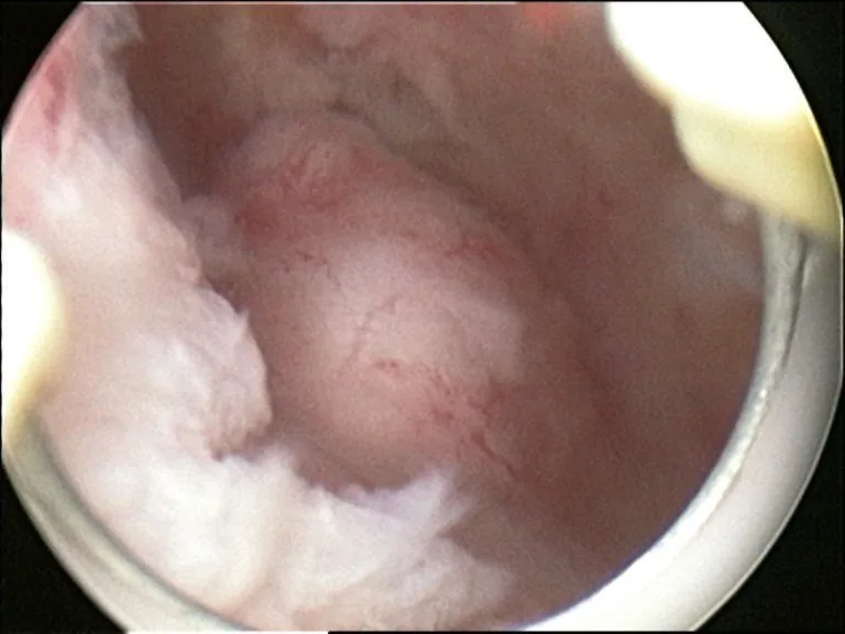 Endoscopy & gross
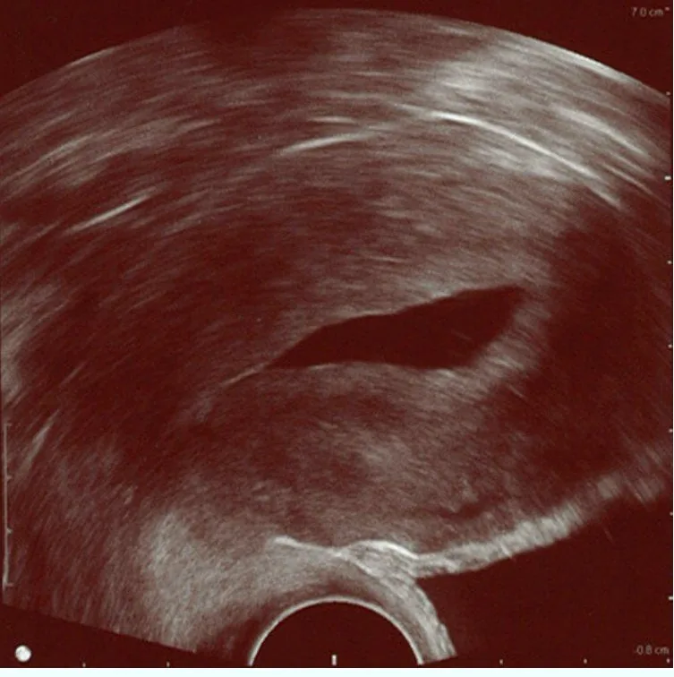 Ultrasound
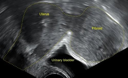 Ultrasound
Definition
- Definition: A fibroid is a benign tumour of smooth muscle cells and connective tissue of the myometrium.
- Histology: The correct histological name is leiomyoma.
- Hormone dependency: Fibroids are oestrogen- and progesterone-dependent, thus growing during reproductive years and shrinking after menopause.
- Frequency: Fibroids are the most common tumour in women overall, symptomatic in about one in four to one in three women over 30.
- Uterus myomatosus: This term describes a uterus enlarged and nodularly deformed by numerous fibroids.
Classification
Special Forms and Features
- Parasitic fibroid: A pedunculated subserosal fibroid with no connection to the uterus is called a parasitic fibroid.
- Red degeneration: This is a haemorrhagic infarction, typically during pregnancy, with acute pain.
- Fibroids in pregnancy: During pregnancy, fibroids can grow and lead to malpresentation, preterm labour, and postpartum atony.
- Postmenopausal growth: Fibroids that start growing after menopause are suspicious and require investigation because a sarcoma may be the underlying cause.
Submucous Types
- FIGO 0: This type describes the completely intracavitary, pedunculated fibroid.
- FIGO 1: This type is defined as the submucous fibroid with less than 50 percent of its volume in the myometrium.
- FIGO 2: This type is defined as the submucous fibroid with 50 percent or more of its volume in the myometrium.
Intramural and Subserous
- FIGO 3: This type is intramural with contact to the endometrium, without bulging into the cavity.
- FIGO 4: This type describes the purely intramural fibroid with no contact to the endometrium or serosa.
- FIGO 5 and 6: FIGO 5 is subserous with at least a 50 percent intramural component, FIGO 6 with less than 50 percent.
- FIGO 7: This type is the pedunculated subserous fibroid.
- FIGO 8: This number designates other locations such as a cervical or parasitic fibroid.
Two-Number Notation
- Meaning: Two numbers, such as 2 to 5, indicate a transmural fibroid extending to both the endometrium and the serosa.
- Order: The first number describes the relationship to the endometrium, the second to the serosa.
Clinical features
Symptoms by Location
- Bleeding disorders: These are primarily caused by submucosal and intramural fibroids near the cavity.
- Cause of bleeding: The reason is the enlarged and less effectively contracting endometrial surface over the nodule.
- Pressure symptoms: Pressure on the bladder and rectum is mainly caused by a large subserosal or near-cervical fibroid.
- Pain development: Pain arises from torsion of a pedunculated fibroid, from necrosis with increasing size, and as dysmenorrhoea.
- Fertility: A fibroid can also be a cause of sterility and miscarriages if it deforms the cavity.
More facts from the study questions
- Fibroids are hormone-dependent tumours that grow during the reproductive years.
- The cause of the heavy bleeding is the enlarged endometrial surface over the fibroid nodule.
- Additionally, the contractility of the myometrium is impaired in this area.
- Subserous fibroids grow on the outside of the uterus and can press on adjacent organs due to their size.
- Typical symptoms are pollakiuria or constipation.
- A fibroid that grows after the menopause is highly suspicious for malignant degeneration.
- Red degeneration is a special form of fibroid necrosis caused by haemorrhagic infarction.
- It characteristically occurs during pregnancy and is associated with acute pain.
- The purpose of the FIGO classification is to describe the location of the fibroid.
- FIGO 4 denotes the classic intramural fibroid.
Diagnosis
History and Examination
- History taking covers bleeding history including amount and duration, pain, pressure symptoms, desire for children and signs of anaemia.
- The bimanual examination typically reveals an enlarged, firm, nodularly contoured and mostly mobile uterus.
Ultrasound
- A fibroid appears as a roundish, hypoechoic, well-defined nodule with circular peripheral vascularisation.
- With a large uterus, one switches to the transabdominal approach, because the organ can no longer be fully visualised transvaginally.
- Documentation includes number, size in three planes, FIGO type and the relationship to the cavum and serosa.
- In case of calcification, this results in a posterior acoustic shadow, which makes assessment more difficult.
Further Procedures
- Hydrosonography with saline solution helps to differentiate submucosal from intramural fibroids and from a polyp.
- In case of abnormal bleeding and risk factors, this includes histological evaluation of the endometrium, because a fibroid and hyperplasia can coexist.
What Raises Suspicion
- Clinical signs: Suspicious signs are rapid growth progression, growth after menopause, pain, and bleeding.
- Sonography: Sonographic indicators are inhomogeneity with necrotic zones, an indistinct border, and disordered, strong central vascularisation.
- Imaging: The crucial statement on imaging is: neither ultrasound nor MRI can reliably rule out a sarcoma.
- Laboratory tests: In terms of laboratory chemistry, there is no useful marker.
Typical imaging findings
- Ultrasound: A rounded hypoechoic mass with concentric internal structure bulges from the wall into the cavity and displaces the endometrial line to the opposite side. Its margin is smooth and separable from the myometrium.
- Ultrasound: A large, rounded mass of about nine centimetres arises from the uterus. Its internal structure is whorled and heterogeneous, with streaky shadowing at the lateral edges; calipers span the nodule.
Keep learning in the app
Further reading (selection)
Cross-references
Note: Learning content from the GynFuchs app (flashcards, exam questions, image cases) for medical education – not a treatment recommendation and no substitute for diagnosis or treatment decisions in individual cases. Treatment and management are deliberately not covered on this page.