Uterine sarcoma (leiomyosarcoma)
Exam relevance: in 7 of 197 board exam reports · rank 88
- Specialty
- Gynaecology · Uterus & endometrium
- Images
- Endoscopy & gross 2
- Exam relevance
- 7 of 197 reports · rank 88
- In the app
- 1 flashcards · GynFuchs
- Last updated
- 10/2026 · Dr. Pascal Bafteh
Contents
Images (2)
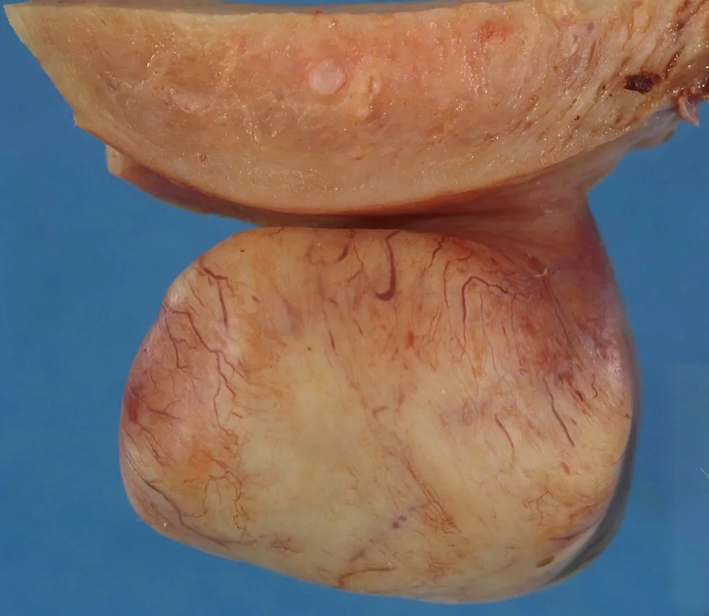 Endoscopy & gross
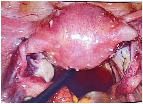 Endoscopy & gross
Definition
- Uterine sarcomas are rare malignant mesenchymal tumours of the uterus; leiomyosarcoma arises from smooth muscle cells or their mesenchymal precursors.
Classification
- Leiomyosarcoma is the most common subtype; others are low-grade and high-grade endometrial stromal sarcoma, undifferentiated uterine sarcoma and adenosarcoma.
- Carcinosarcomas are of epithelial origin and are no longer regarded as uterine sarcomas.
Occurrence & epidemiology
Risk
- Sarcoma risk: A finding clinically classified as a fibroid conceals a sarcoma in the order of approximately 0.2 to 0.3 percent.
- Risk factors: The risk increases with age and is significantly higher after menopause.
- Entities: The most common entity is leiomyosarcoma, in addition to endometrial stromal sarcoma and undifferentiated sarcoma.
- Origin: A leiomyosarcoma usually arises de novo and not from a pre-existing fibroid.
Epidemiology and risk factors
- Uterine sarcomas account for about 3–7% of all uterine malignancies.
- Incidence is higher from the age of 50 than in younger women and about twice as high in women of Afro-Caribbean descent as in white women.
Clinical features
- Symptoms are often non-specific: lower abdominal or pelvic pain, abdominal distension and, most commonly, abnormal vaginal bleeding.
- A rapidly growing presumed fibroid in a peri- or postmenopausal woman is suspicious; malignancy is often only recognised on histology.
Histology
- Leiomyosarcoma is distinguished from leiomyoma morphologically by mitoses, atypia and necrosis.
- Spindle cells in intersecting fascicles with co-expression of SMA, desmin and h-caldesmon are typical.
Diagnosis
What Raises Suspicion
- Clinical signs: Suspicious signs are rapid growth progression, growth after menopause, pain, and bleeding.
- Sonography: Sonographic indicators are inhomogeneity with necrotic zones, an indistinct border, and disordered, strong central vascularisation.
- Imaging: The crucial statement on imaging is: neither ultrasound nor MRI can reliably rule out a sarcoma.
- Laboratory tests: In terms of laboratory chemistry, there is no useful marker.
- Ultrasound and MRI cannot yet reliably distinguish a leiomyoma from a sarcoma; the diagnosis is histological.
- FIGO staging for uterine sarcomas has been adapted to the histological subtype.
Keep learning in the app
Further reading (selection)
Cross-references
Note: Learning content from the GynFuchs app (flashcards, exam questions, image cases) for medical education – not a treatment recommendation and no substitute for diagnosis or treatment decisions in individual cases. Treatment and management are deliberately not covered on this page.