Hormone receptor-positive breast cancer (luminal A and B)
Exam relevance: in 14 of 197 board exam reports · rank 55
- Specialty
- Gynaecology · Breast
- Images
- Histology & cytology 2
- Exam relevance
- 14 of 197 reports · rank 55
- Last updated
- 10/2026 · Dr. Pascal Bafteh
Contents
Images (2)
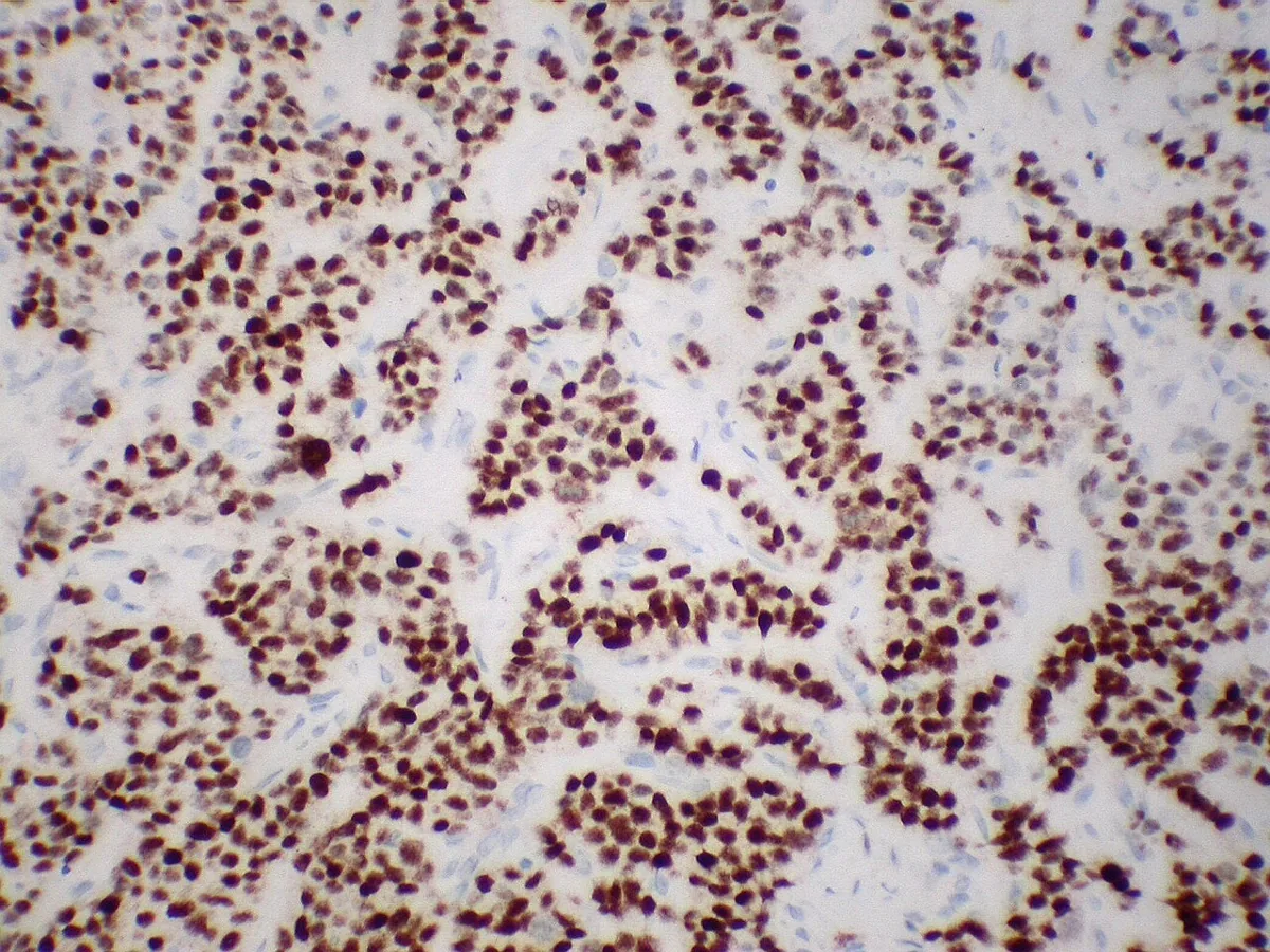 Histology & cytology
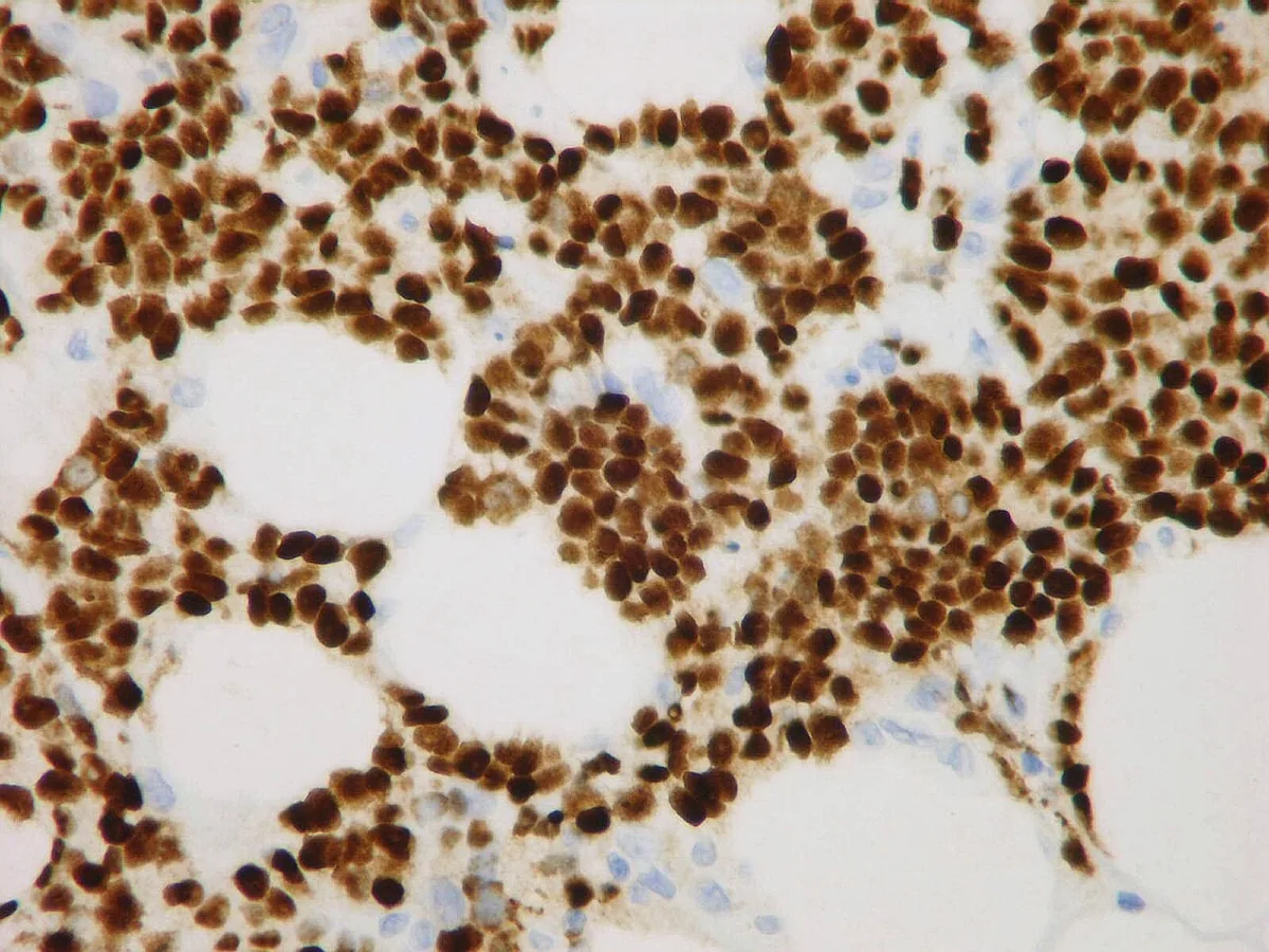 Histology & cytology
Definition
- Hormone receptor-positive breast cancer with oestrogen and/or progesterone receptors, corresponding to the luminal A and luminal B molecular subtypes.
Classification
- Luminal A tumours are grade 1 or 2, oestrogen and progesterone receptor-positive, HER2-negative and have low Ki-67.
- Luminal B tumours are oestrogen receptor positive, often have a high Ki-67 fraction and may be HER2 positive or negative.
Occurrence & epidemiology
Epidemiology
- Most breast cancers are luminal A; about 70% are hormone receptor-positive and HER2-negative.
- Most oestrogen receptor-positive tumours are also progesterone receptor-positive; overall, about 70% of all breast cancers are progesterone receptor-positive.
Aetiopathogenesis
Aetiology and pathogenesis
- Oestrogen and progesterone receptors are nuclear hormone receptors that promote DNA replication and cell division when their hormones bind, so oestrogen and progesterone can fuel tumour growth.
- Early menarche, late menopause and older age at first pregnancy increase breast cancer risk.
Clinical features
- Luminal A and B tumours generally grow more slowly than HER2-enriched and triple-negative (basal-like) tumours.
Diagnosis
- Breast cancers are usually tested for oestrogen and progesterone receptors and HER2, often also for the Ki-67 protein.
- Most guidelines regard immunohistochemical staining of 1% or more as oestrogen receptor-positive; ER-low tumours with 1–10% expression (about 2–7% of cases) resemble oestrogen receptor-negative tumours at the molecular level.
- Ki-67 is found only in dividing cells, and a high proportion of Ki-67-positive tumour cells indicates rapid division.
- Hormone receptor status can change over time.
Keep learning in the app
Further reading (selection)
Cross-references
More topics: Breast
Note: Learning content from the GynFuchs app (flashcards, exam questions, image cases) for medical education – not a treatment recommendation and no substitute for diagnosis or treatment decisions in individual cases. Treatment and management are deliberately not covered on this page.