HER2-positive breast cancer
Exam relevance: in 38 of 197 board exam reports · rank 15
- Specialty
- Gynaecology · Breast
- Images
- Ultrasound 2
- Exam relevance
- 38 of 197 reports · rank 15
- In the app
- 1 flashcards · GynFuchs
- Last updated
- 10/2026 · Dr. Pascal Bafteh
Contents
Images (2)
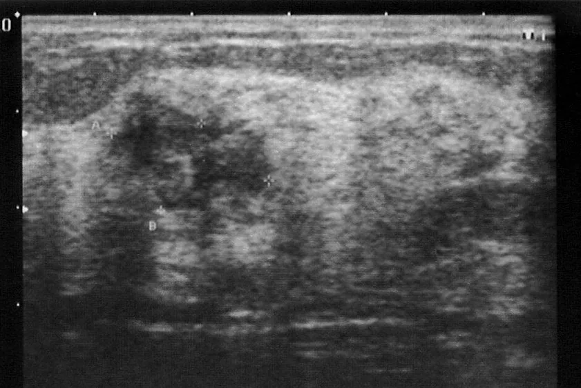 Ultrasound
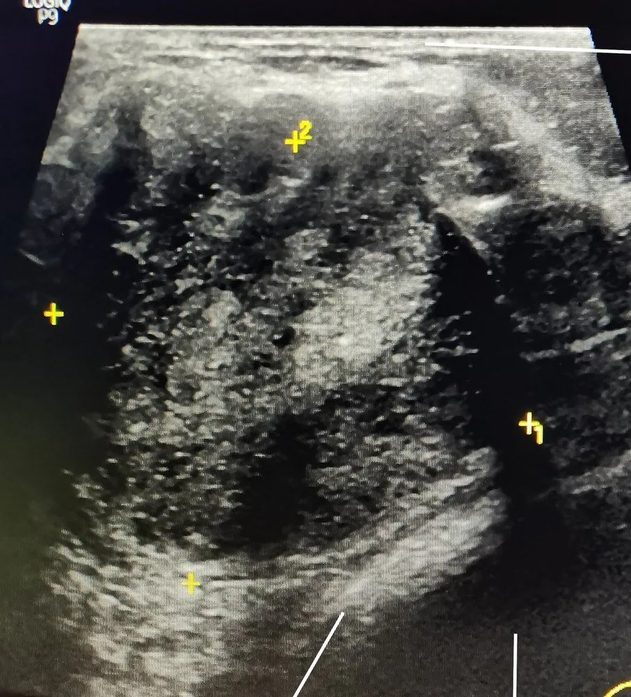 Ultrasound
Definition
- Breast cancer with amplification of the HER2 gene and/or overexpression of the HER2 protein, a transmembrane tyrosine kinase receptor of the HER family of growth factor receptors (HER1–4).
Classification
Molecular subtypes
- Luminal A: A Luminal A carcinoma is hormone receptor-positive, HER2-negative, well-differentiated with a low Ki-67.
- Luminal B: A Luminal B carcinoma is hormone receptor-positive with a high Ki-67 or poorer grade, HER2 negative or positive.
- Triple-negative: In triple-negative breast cancer: oestrogen receptor, progesterone receptor, and HER2 are all negative.
- HER2-positive tumours may be hormone receptor-positive (luminal B subtype) or hormone receptor-negative; the latter form the HER2-positive (HER2-enriched) subtype.
- HER2-low denotes tumours with an immunohistochemical score of 1+ or 2+ without amplification on in situ hybridisation; they contain some HER2 protein but not enough to be classified as HER2-positive.
- HER2-low is a heterogeneous group, and whether it constitutes a distinct entity remains controversial.
Occurrence & epidemiology
Epidemiology
- About 15–20% of breast cancers are HER2-positive, about 40–60% are HER2-low, and 20–45% show no HER2 expression (score 0).
Aetiopathogenesis
Aetiology and pathogenesis
- The HER2 gene lies on chromosome 17 (band q21); tumour tissue may have up to 25–50 gene copies, up to a 40- to 100-fold increase in protein and up to 2 million receptors on the cell surface.
- HER2 itself binds no ligand but is activated by homo- or heterodimerisation with the ligand-bound receptors HER1, HER3 and HER4, mainly switching on the MAPK and PI3K signalling pathways.
- These pathways promote cell proliferation, invasion and angiogenesis and protect cells against apoptosis.
Clinical features
More facts from the study questions
- The Elston and Ellis grading system is an established system for assessing the aggressiveness of breast carcinoma.
- It is based on the histological evaluation of three criteria: tubule formation, nuclear pleomorphism and mitotic rate.
- The definition of triple-negative breast carcinoma is the lack of expression of all three relevant receptors.
- It is oestrogen receptor-negative, progesterone receptor-negative and HER2-negative.
- HER2 status is primarily determined by immunohistochemistry (IHC).
- Invasive lobular carcinoma is characterised by diffuse growth and is difficult to delineate.
- HER2 positivity gives the tumour a more aggressive behaviour, with higher recurrence rates and shorter disease-free and overall survival.
- HER2-positive carcinomas tend to grow quickly, faster than tumours with less HER2 (HER2-negative or HER2-low).
Diagnosis
- HER2 status is determined on tumour tissue; only immunohistochemistry for protein expression and in situ hybridisation for gene status are clinically validated.
- A tumour is HER2-positive with an immunohistochemical score of 3+ or a score of 2+ with amplification demonstrated by in situ hybridisation; 2+ without amplification, 1+ and 0 count as HER2-negative.
- Pre-analytical and analytical factors such as tissue fixation, processing, staining technique and scoring criteria largely determine the reliability of the result.
Keep learning in the app
Further reading (selection)
Cross-references
More topics: Breast
Note: Learning content from the GynFuchs app (flashcards, exam questions, image cases) for medical education – not a treatment recommendation and no substitute for diagnosis or treatment decisions in individual cases. Treatment and management are deliberately not covered on this page.