Inflammatory breast cancer
Exam relevance: in 9 of 197 board exam reports · rank 75
- Specialty
- Gynaecology · Breast
- Images
- Clinical 2 · Ultrasound 4
- Exam relevance
- 9 of 197 reports · rank 75
- In the app
- 1 flashcards · GynFuchs
- Last updated
- 10/2026 · Dr. Pascal Bafteh
Contents
Images (6)
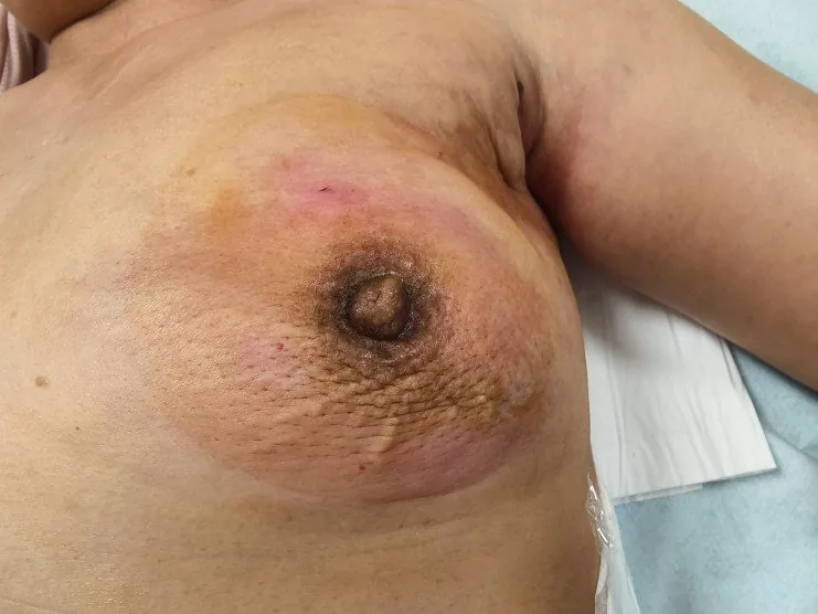
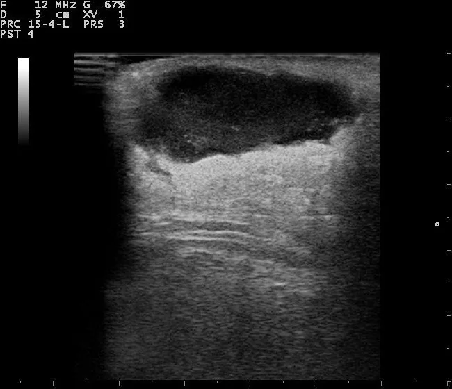 Ultrasound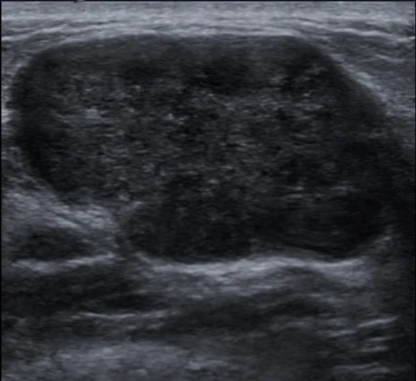
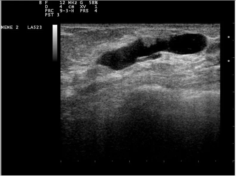 Ultrasound
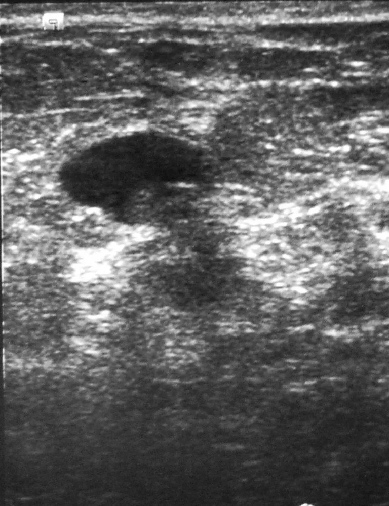 Ultrasound
 Ultrasound
Definition
- Clinicopathological entity of breast cancer with erythema and oedema over at least one third of the breast, classified as T4d in the TNM system.
Classification
- Diagnostic criteria are a rapid onset of erythema, oedema, peau d'orange or warmth with or without a palpable mass, a history of no more than six months, erythema of at least one third of the breast and histological confirmation of invasive carcinoma.
- The primary form develops in a previously normal breast, the secondary form as inflammatory skin change in a breast with known invasive cancer.
Occurrence & epidemiology
Epidemiology
- Inflammatory breast cancer accounts for about 1–5% of breast cancers in the United States, depending on the source, but for about 7% of breast cancer deaths.
- It is often diagnosed at a younger age and is more common and diagnosed earlier in Black women than in White women.
Aetiopathogenesis
Aetiology and pathogenesis
- The cause is unknown; the disease is more common in women with obesity.
- The typical picture arises because tumour cells block the lymphatic vessels of the skin; most tumours are invasive ductal carcinomas and often hormone receptor-negative.
Clinical features
Distinction from inflammation
- Inflammatory breast cancer presents with diffuse erythema over at least one-third of the breast with oedema and peau d'orange, characteristically without fever.
- Inflammatory carcinoma is confirmed by a punch biopsy of the skin showing lymphatic vessel invasion, as a discrete mass is often absent.
- Paget's disease of the nipple is an intraepidermal spread of carcinoma cells and thus a sign of cancer, not a dermatosis.
- Symptoms develop rapidly; the breast is red and swollen, enlarges rapidly and shows peau d'orange, and there is usually no palpable lump.
- Further features are heaviness, burning or tenderness, an inverted nipple and enlarged lymph nodes in the axilla or near the collarbone.
- At diagnosis the disease is at least stage IIIB; most patients have locoregional disease, and up to 40% already have distant metastases.
Diagnosis
Typical imaging findings
- Clinical finding: The breast is diffusely red over a wide area, clearly enlarged and warm. The skin is thickened with coarse pitting — the peau d'orange appearance; the redness involves more than a third of the breast and there is no circumscribed mass.
- Mammography frequently shows skin thickening (84–93%), increased density (93%) and axillary adenopathy (24%), and ultrasound shows skin thickening, hypoechoic non-mass abnormalities, calcifications and nodal disease.
- The diagnosis is confirmed by core needle biopsy, supplemented by a skin punch biopsy from the most affected area; tumour cell emboli in dermal lymphatics are typical but not required for the diagnosis.
Keep learning in the app
Further reading (selection)
Cross-references
More topics: Breast
Note: Learning content from the GynFuchs app (flashcards, exam questions, image cases) for medical education – not a treatment recommendation and no substitute for diagnosis or treatment decisions in individual cases. Treatment and management are deliberately not covered on this page.