Lobular breast cancer and lobular neoplasia
Exam relevance: in 5 of 197 board exam reports · rank 101
- Specialty
- Gynaecology · Breast
- Images
- Histology & cytology 3
- Exam relevance
- 5 of 197 reports · rank 101
- Last updated
- 10/2026 · Dr. Pascal Bafteh
Contents
Images (3)
 Histology & cytology
 Histology & cytology
 Histology & cytology
Definition
- Lobular breast lesions consist of small, noncohesive tumour cells arising from the lobules or the terminal duct lobular unit (TDLU).
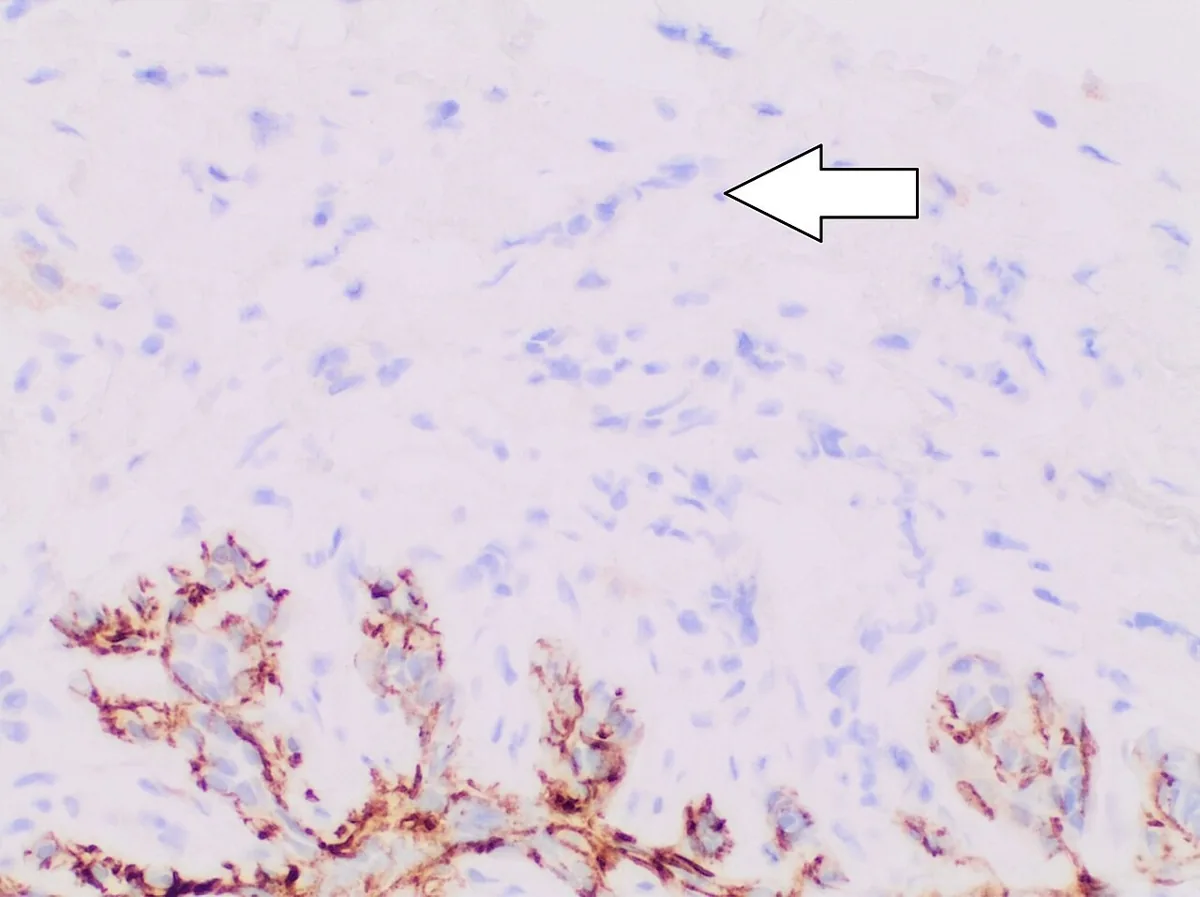
- The molecular hallmark is loss of E-cadherin, caused by a CDH1 mutation in more than 90% of invasive lobular carcinomas.
Classification
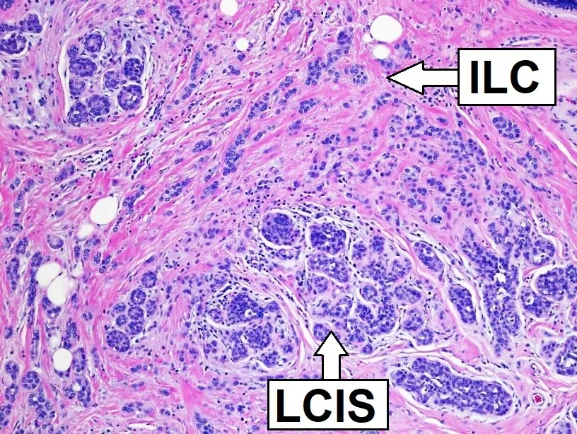
- Precursor lesions are atypical lobular hyperplasia (ALH) and lobular carcinoma in situ (LCIS): ALH involves and distends less than 50% of the acini of a TDLU, LCIS more than 50%.
- Besides classic invasive lobular carcinoma, the WHO classification lists solid, alveolar, pleomorphic and tubulolobular variants. Solid forms are more often high grade and associated with worse overall survival, while pleomorphic forms more often show HER2 amplification or a triple-negative profile.
Occurrence & epidemiology
Epidemiology
- Invasive lobular carcinoma is the second most common type of invasive breast cancer, accounting for 10–15% of cases.
- It tends to occur in older women and is over-represented among bilateral breast cancers.
Clinical features
- Because of its diffuse growth the carcinoma often forms no palpable lump; clinical and radiological findings are subtle, and diagnosis is often delayed.
- Multifocality, multicentricity and bilaterality are more common, and metastases also occur in the gastrointestinal tract, peritoneum, retroperitoneum, ovaries, meninges and orbits.
Histology
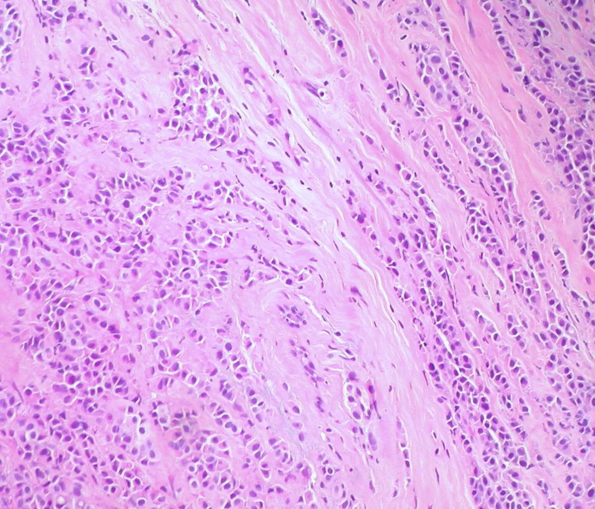
- The tumour cells lie singly or in single-file cords within fibrous stroma with little desmoplastic reaction and often encircle normal ducts in a targetoid fashion.
- Invasive lobular carcinoma is almost always oestrogen and progesterone receptor-positive, and HER2 overexpression or amplification is rare (3–13%).
- Lobular neoplasia is found alongside in about half of cases.
Diagnosis
- On mammography the carcinoma is often subtle; it most commonly presents as a spiculated mass or architectural distortion, and up to 30% are mammographically occult.
- The sensitivity of mammography for lobular carcinoma is 34–92% (ductal 63–98%), and that of MRI is 83–100%, above 95% in most studies.
Keep learning in the app
Further reading (selection)
- StatPearls: Invasive Lobular Carcinoma (NCBI Bookshelf)
- Lobular Breast Cancer: Histomorphology and Different Concepts of a Special Spectrum of Tumors (Cancers (Basel) 2021, PubMed Central)
- Invasive Lobular Carcinoma: A Review of Imaging Modalities with Special Focus on Pathology Concordance (Healthcare (Basel) 2023, PubMed Central)
Cross-references
Note: Learning content from the GynFuchs app (flashcards, exam questions, image cases) for medical education – not a treatment recommendation and no substitute for diagnosis or treatment decisions in individual cases. Treatment and management are deliberately not covered on this page.