Breast abscess
Exam relevance: in 1 of 197 board exam reports · rank 171
- Specialty
- Gynaecology · Breast
- Images
- Ultrasound 1 · Clinical 1
- Exam relevance
- 1 of 197 reports · rank 171
- Last updated
- 10/2026 · Dr. Pascal Bafteh
Contents
Images (2)
 Ultrasound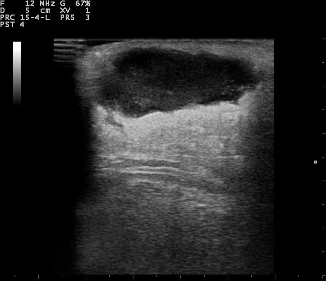
Definition
- Organised collection of inflammatory exudate, or pus, in the breast.
Classification
- Lactational and nonlactational abscesses are distinguished, corresponding broadly to postpartum and non-postpartum disease; they are regarded as distinct entities with their own pathogenesis.
Occurrence & epidemiology
Epidemiology
- Breast infections mostly affect women aged 18 to 50, but abscesses also occur in adolescents, neonates and men.
- Lactational mastitis is common, with a prevalence of about 1–10% among lactating women, whereas an abscess is rare, occurring in about 0.4–0.5% of lactating women in most cohort studies.
- Nonlactational abscesses are seen more often towards the end of the reproductive years.
Aetiopathogenesis
Aetiology and pathogenesis
- An abscess is a complication of both lactational and nonlactational mastitis; during lactation, microorganisms can be transmitted from the infant to the nipple and areola when the epithelial barrier is compromised.
- Lactational abscesses are most often caused by Staphylococcus aureus and streptococci, and MRSA is increasingly seen.
- Nonlactational abscesses are mostly subareolar, are associated with smoking, diabetes and ductal changes and are usually caused by mixed flora; chronic or recurrent abscesses often also involve anaerobes.
Clinical features
- Typical are localised pain, tenderness, swelling and erythema of one breast, frequently with fever, fatigue and flu-like symptoms; the axillary lymph nodes may be reactively enlarged.
- Nonlactational abscesses tend to recur, with reported recurrence rates of 25–50%.
- Particularly in nonlactating women, a serious underlying condition such as malignancy or previously undiagnosed diabetes may be present.
Diagnosis
Typical imaging findings
- Clinical finding: The breast is red and tense; right next to the nipple there is a freshly incised wound with an indwelling drain, and the dressing shows a mix of blood, pus and breast milk.
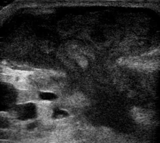
- Ultrasound: The affected area shows an ill-defined hypoechoic zone with internal echoes (debris) and a heterogeneous structure — consistent with a purulent liquefaction within inflamed tissue.
- Ultrasound is the primary imaging modality; the abscess appears as a focal fluid collection that is irregular and hypoechoic, with poorly defined margins and heterogeneous internal echoes.
- Mobile internal debris within the collection helps to distinguish an abscess from an inflammatory phlegmon; scanning with the probe is frequently tender.
- Blood count and CRP can indicate systemic infection or severe inflammation.
Keep learning in the app
Further reading (selection)
Cross-references
More topics: Breast
Note: Learning content from the GynFuchs app (flashcards, exam questions, image cases) for medical education – not a treatment recommendation and no substitute for diagnosis or treatment decisions in individual cases. Treatment and management are deliberately not covered on this page.