Intraductal papilloma
Exam relevance: in 1 of 197 board exam reports · rank 171
- Specialty
- Gynaecology · Breast
- Images
- Clinical 2 · Ultrasound 4
- Exam relevance
- 1 of 197 reports · rank 171
- In the app
- 1 flashcards · GynFuchs
- Last updated
- 10/2026 · Dr. Pascal Bafteh
Contents
Images (6)
 Ultrasound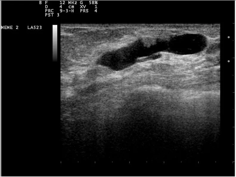
 Ultrasound
 Ultrasound
 Ultrasound
Definition
- Benign intraluminal proliferation within the breast ducts and the most common papillary lesion of the breast.
Classification
- Central papillomas lie in the large ducts behind the nipple and are more common; peripheral papillomas are smaller and often multiple.
- Depending on the extent of atypical proliferation, papillomas with atypical ductal hyperplasia (ADH) or with DCIS are distinguished from papillomas without atypia.
- In the European classification, papilloma fragments on core or vacuum-assisted biopsy are categorised as B3 lesions (uncertain malignant potential) regardless of atypia, because atypia elsewhere in the lesion cannot be excluded.
Occurrence & epidemiology
Epidemiology
- Papillomas occur mostly in perimenopausal women aged 30 to 50 years; intraductal papillomas account for approximately 20% of B3 lesions.
Clinical features
Pathological nipple discharge
- A suspicious discharge is one that is unilateral, spontaneous, bloody, and from a single duct orifice.
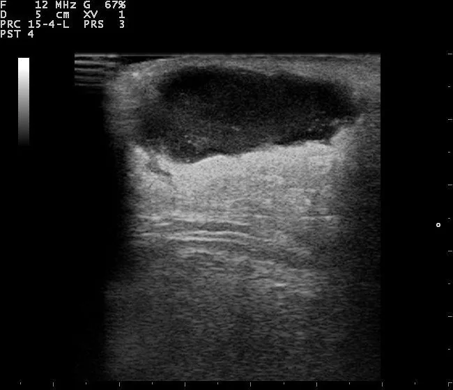
- On ultrasound, in cases of suspicious discharge, one looks for a dilated duct with an intraluminal mass, such as an intraductal papilloma.
- Central papillomas often cause serous or bloody nipple discharge; intraductal papilloma is the most common cause of bloody nipple discharge without a breast mass.
- Peripheral papillomas are usually asymptomatic and may be associated with microcalcifications detected on mammography.
Histology
- Histology shows arborising fibrovascular cores within a dilated duct, lined by myoepithelial cells and covered by luminal epithelial cells; this two-cell-type pattern is the hallmark of benign papillary lesions.
- Sclerosis and squamous, apocrine, mucinous or chondroid metaplasia may be present; epithelial proliferations such as ADH, atypical lobular hyperplasia or DCIS are more common in peripheral papillomas.
- The risk that DCIS or invasive carcinoma is found in subsequently obtained tissue after core biopsy of a papilloma without atypia is given as 2–3%; across studies this upgrade rate ranges from 0 to 16%.
Diagnosis
Typical imaging findings
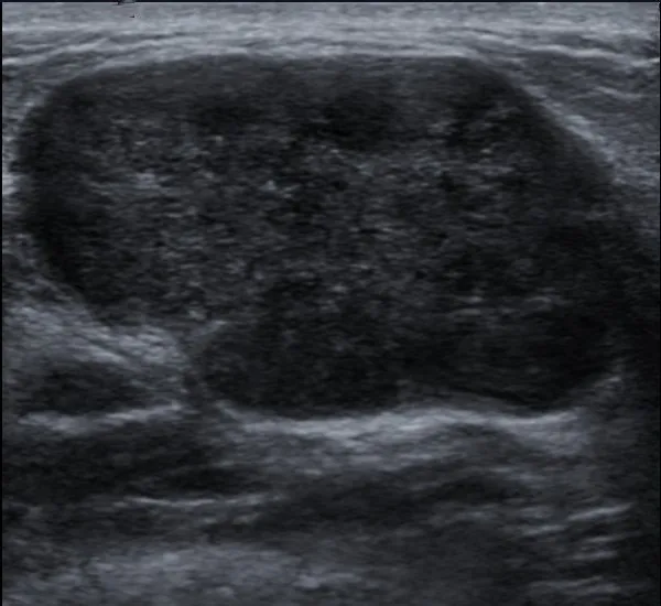
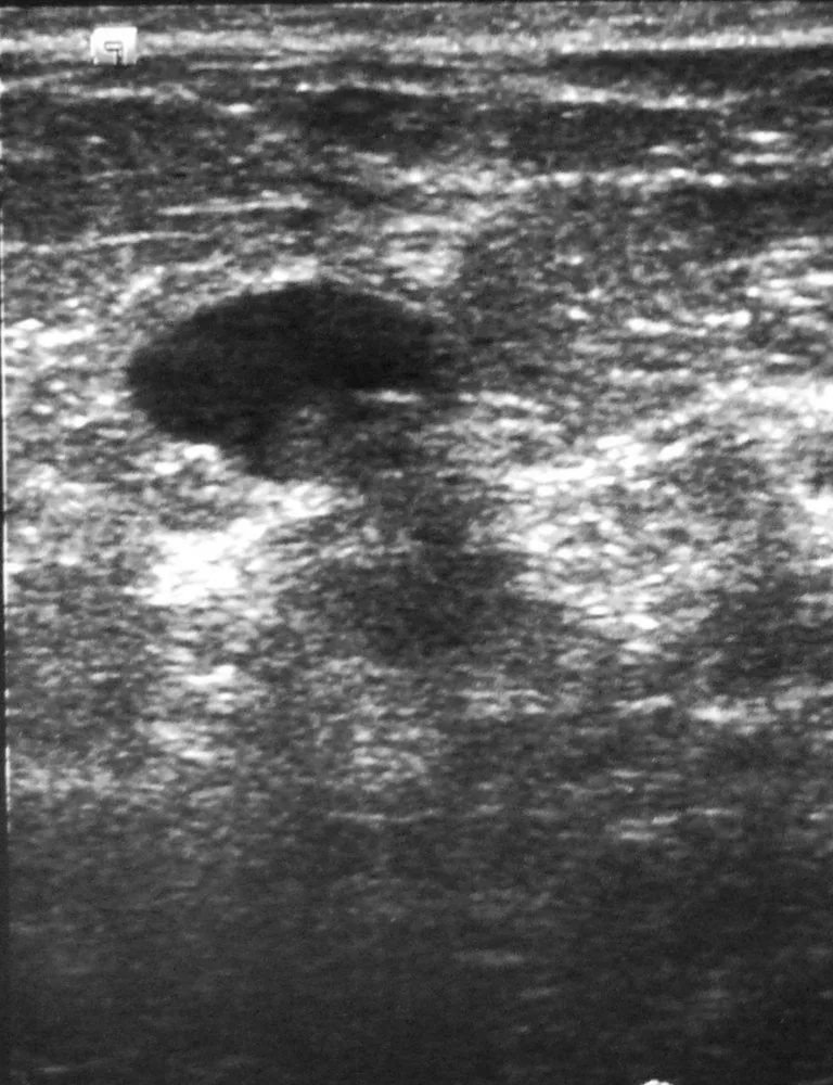
- Ultrasound: Behind the nipple a duct is anechoically dilated. A solid hypoechoic plug sits within its lumen, broadly attached to the wall; the duct can be followed on both sides of it as a fluid-filled tube.
- On mammography papillomas, particularly small ones, are often unremarkable; larger lesions may appear as well-defined round or oval opacities with or without microcalcifications.
- On ultrasound the papilloma typically appears as a hypoechoic, well-defined lesion filling a duct or partly outlined by fluid, sometimes with a vascular stalk.
- Galactography shows filling defects in the affected duct; the diagnosis is confirmed by ultrasound-guided biopsy.
Keep learning in the app
Further reading (selection)
Cross-references
More topics: Breast
Note: Learning content from the GynFuchs app (flashcards, exam questions, image cases) for medical education – not a treatment recommendation and no substitute for diagnosis or treatment decisions in individual cases. Treatment and management are deliberately not covered on this page.