Vulvar Paget disease
Exam relevance: in 1 of 197 board exam reports · rank 171
- Specialty
- Gynaecology · Vulva & vagina
- Images
- Histology & cytology 1 · Clinical 1
- Exam relevance
- 1 of 197 reports · rank 171
- In the app
- 1 flashcards · GynFuchs
- Last updated
- 10/2026 · Dr. Pascal Bafteh
Contents
Images (2)
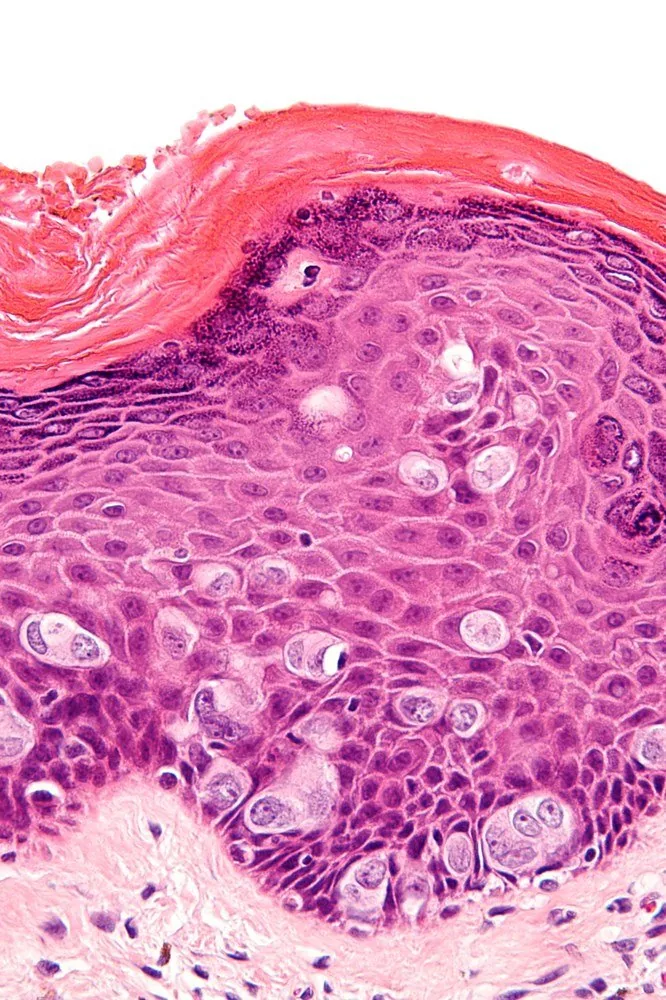 Histology & cytology
Definition
Paget's Disease of the Vulva
- Histology: Histologically, one finds large, pale, vacuolated Paget cells, singly and in nests within the epidermis.
- Classification: The disease is classified into a primary cutaneous form and a secondary form resulting from the spread of an anorectal or urothelial carcinoma.
- Work-up: A complete work-up therefore includes examination of the anorectum and urinary tract, as well as a search for an invasive component.
- Extramammary Paget disease is a rare cutaneous adenocarcinoma arising in skin regions with a high density of apocrine glands, most frequently the vulva.
Classification
- The primary type arises within the epidermis, thought to result from malignant transformation of a cell in the underlying adnexal structures.
- The secondary type results from epidermotropic spread of an underlying visceral carcinoma, most commonly of anorectal or urothelial origin; in previous cohorts it accounted for about 11–18 % of cases.
Occurrence & epidemiology
Epidemiology
- Mainly older adults are affected, typically between 50 and 80 years of age.
- About 10–30 % of patients have an associated internal malignancy, most commonly of the colon, breast or genitourinary tract.
Clinical features
- The most common symptom is itching; lesions mostly present as erythema or red plaques, often with erosions, scaling or hypo- or hyperpigmentation on the surface.
- The lesions expand slowly and progress chronically; because the picture mimics benign infectious or inflammatory dermatoses, the diagnosis is frequently delayed.
Histology
- Paget cells lie scattered or in nests within the epidermis; they are large, round to oval and have abundant pale cytoplasm and large hyperchromatic nuclei.
- The primary type typically expresses CK7 and TRPS1, but GCDFP-15 in only about 68 % of cases; secondary disease of colorectal origin usually expresses CK20, CDX2 and SATB2, urothelial disease GATA3, p63 and uroplakin II/III.
- The profiles overlap: CK20 is found in about 22 % of primary and CK7 in about 81 % of secondary cases; S100 is not expressed in either type.
Diagnosis
- The diagnosis is confirmed by skin biopsy; a panel of immunohistochemical markers rather than a single marker is used to distinguish the primary from the secondary type.
Keep learning in the app
Further reading (selection)
- StatPearls: Extramammary Paget Disease (NCBI Bookshelf)
- Diagnostic Algorithm for Secondary Extramammary Paget Disease from Institutional Cases and Literature Review (Cancers 2025, PubMed Central)
- Clinicopathological Analysis of 40 Cases of Extramammary Paget Disease: A Retrospective Study (Clin Cosmet Investig Dermatol 2026, PubMed Central)
- DocCheck Flexikon, Morbus Paget
Cross-references
Note: Learning content from the GynFuchs app (flashcards, exam questions, image cases) for medical education – not a treatment recommendation and no substitute for diagnosis or treatment decisions in individual cases. Treatment and management are deliberately not covered on this page.