Vulvar lichen sclerosus
Exam relevance: in 10 of 197 board exam reports · rank 66
- Synonyms
- lichen sclerosus
- Specialty
- Gynaecology · Vulva & vagina
- Images
- Clinical 1 · Histology & cytology 1
- Exam relevance
- 10 of 197 reports · rank 66
- In the app
- 1 flashcards · GynFuchs
- Last updated
- 10/2026 · Dr. Pascal Bafteh
Contents
Images (2)
 Histology & cytology
Clinical features
Clinical Presentation
- Definition: Lichen sclerosus is a chronic inflammatory, likely autoimmune-mediated dermatosis.
- Age of onset: The disease has two peaks of onset, namely prepubertal and postmenopausal.
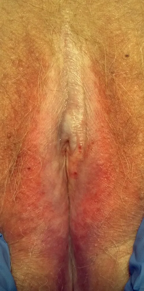
- Skin findings: Typical findings are ivory-white, atrophic, cigarette paper-like skin with itching, burning and fissures.
- Distribution pattern: The characteristic distribution pattern is butterfly- or figure-of-eight-shaped around the vulva and anus.
- Loss of architecture: Advanced findings include effacement of the labia minora, fusion of the clitoral hood, synechiae and introital stenosis.
- Associations: It is frequently associated with other autoimmune diseases, especially Hashimoto's thyroiditis and vitiligo.
Diagnosis
- Establishing the diagnosis: The diagnosis is usually made clinically.
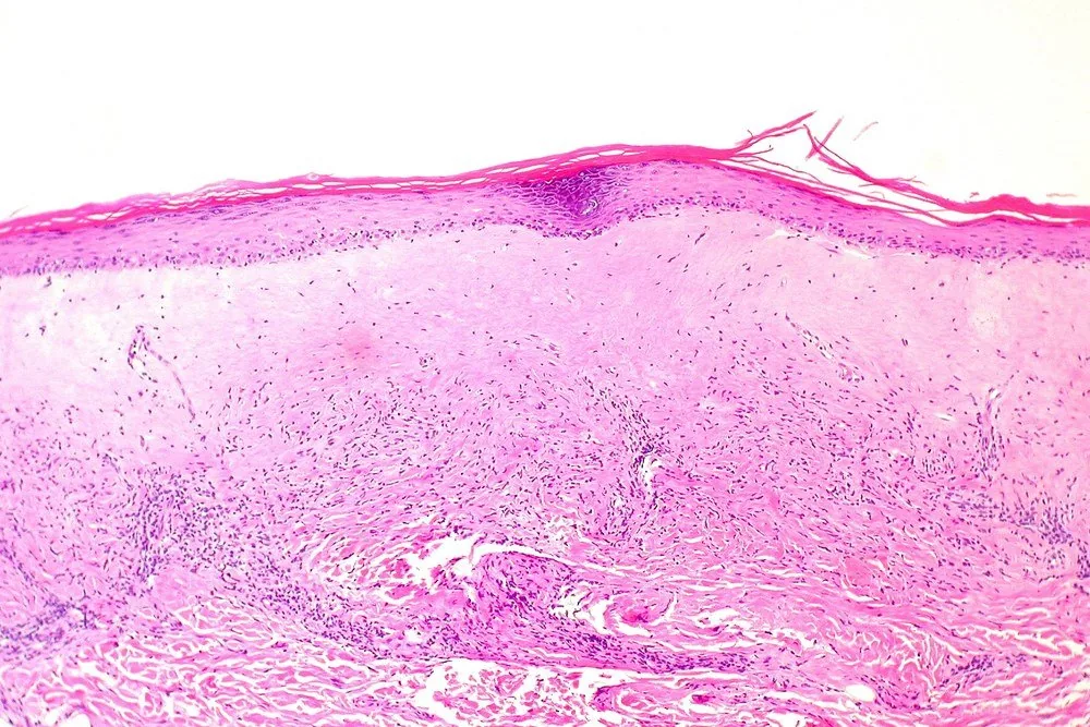
- Histology: Histologically, one finds epidermal atrophy, a broad band of homogenised collagen in the upper dermis, and an underlying lymphocytic infiltrate.
Typical imaging findings
- Clinical finding: Around the clitoris and along the labia the skin is ivory-white, thin and parchment-like; the labia minora are effaced and fused with the labia majora. The inner aspects are reddened, with small fissures and scratch marks.
Keep learning in the app
Further reading (selection)
Cross-references
Note: Learning content from the GynFuchs app (flashcards, exam questions, image cases) for medical education – not a treatment recommendation and no substitute for diagnosis or treatment decisions in individual cases. Treatment and management are deliberately not covered on this page.