Anencephaly
Exam relevance: in 4 of 197 board exam reports · rank 111
- Specialty
- Obstetrics · Fetus & prenatal medicine
- Images
- Ultrasound 2
- Exam relevance
- 4 of 197 reports · rank 111
- Last updated
- 10/2026 · Dr. Pascal Bafteh
Contents
Images (2)
 Ultrasound
 Ultrasound
Definition
- Anencephaly is a developmental disorder in which the skull vault is absent and most or all of the brain tissue is lacking; the brainstem, cerebellum and diencephalon are usually present.
- It belongs to the neural tube defects and results from failure of closure of the rostral end of the neural tube; failure of the caudal end causes spina bifida.
Classification
- A distinction is made between meroacrania (foramen magnum not involved), holoacrania (defect extending beyond the foramen magnum) and holoacrania with rachischisis when spina bifida coexists.
Aetiopathogenesis
Aetiology and risk factors
- The neural tube forms in the fourth week of development; the cranial neuropore closes around day 25, the caudal neuropore about two days later.
- If the rostral neuropore remains open, exencephaly develops first; without a protective calvarium the brain tissue degenerates while the brainstem is preserved (acrania–exencephaly–anencephaly sequence).
- The aetiology is multifactorial; one of the most important nutritional factors is folate deficiency, together with antiepileptic agents and excess vitamin A intake via folate antagonism; valproate is considered the most teratogenic.
- Further maternal factors are diabetes, obesity, exposure to toxins, a positive family history of neural tube defects and high fever or hyperthermia (sauna, hot tub) in early pregnancy.
Clinical features
Clinical features and complications
- Anencephaly is incompatible with life: mortality is 100 per cent, in utero or within hours to days after birth.
- Additional structural anomalies are present in 12 to 25 per cent.
Diagnosis
Typical imaging findings
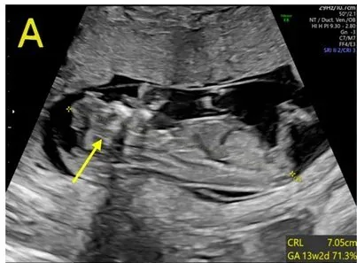
- Ultrasound: In the sagittal plane the trunk is normally developed, but above the orbits the echogenic outline of the vault is missing. Brain tissue appears irregular and without a surrounding bony shell.
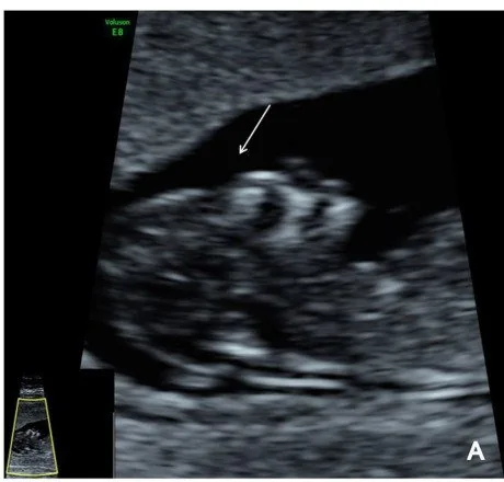
- Ultrasound: Above the fetal facial plane the bony cranial vault is completely absent. Brain tissue lies unprotected on the skull base and floats in the amniotic fluid; the head contour ends abruptly above the orbits.
- On first-trimester ultrasound anencephaly is now detected in virtually all cases; transvaginal imaging shows acrania and a variable degree of brain degeneration.
- Gestational age derived from crown–rump length is on average about two weeks less than expected in affected fetuses.
- In anencephaly the cerebral hemispheres are replaced by masses of connective and vascular tissue.
- Raised maternal serum alpha-fetoprotein suggests open spina bifida, anencephaly or an abdominal wall defect.
Keep learning in the app
Further reading (selection)
Cross-references
More topics: Fetus & prenatal medicine
Note: Learning content from the GynFuchs app (flashcards, exam questions, image cases) for medical education – not a treatment recommendation and no substitute for diagnosis or treatment decisions in individual cases. Treatment and management are deliberately not covered on this page.