Fetal anaemia
Exam relevance: in 9 of 197 board exam reports · rank 75
- Specialty
- Obstetrics · Fetus & prenatal medicine
- Images
- Ultrasound 1 · CTG 4 · Clinical 1
- Exam relevance
- 9 of 197 reports · rank 75
- In the app
- 2 flashcards · GynFuchs
- Last updated
- 10/2026 · Dr. Pascal Bafteh
Contents
Images (6)
 Ultrasound
 CTG
CTG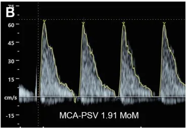
 CTG
CTG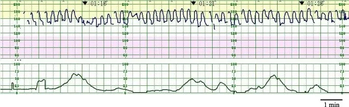 CTG
CTG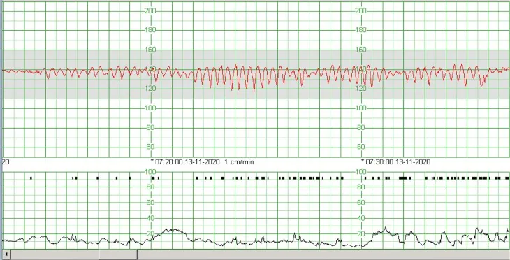 CTG
CTGDefinition
- Fetal anaemia is a reduction of fetal haemoglobin below the value expected for gestational age.
Aetiopathogenesis
Causes
- The leading cause is severe fetal anaemia.
- Classic triggers are parvovirus B19, fetomaternal haemorrhage.
- A pseudo-sinusoidal pattern follows opioids such as pethidine and is shorter, less regular and interrupted by accelerations.
Aetiology
- The most important immune cause is alloimmunisation: a Rh-negative mother forms antibodies after exposure to Rh-positive fetal red cells, which in subsequent pregnancies cross the placenta and destroy fetal red cells.
- Antibodies against other blood group systems such as Kell, Duffy, Kidd or Cc and Ee can also cause haemolytic disease; anti-Kell antibodies additionally suppress erythropoiesis in the bone marrow.
- Parvovirus B19 infects fetal tissues expressing the P antigen receptor, including haematopoietic cells; fetuses in the first and second trimesters are particularly at risk because red cell progenitors develop mainly during this period.
Clinical features
Clinical features and complications
- Fetal anaemia can lead to hypoalbuminaemia, high-output heart failure, hydrops fetalis and fetal death; ultrasound signs of hydrops are ascites, skin oedema and pleural and pericardial effusions.
- After birth, in haemolytic causes, jaundice and hyperbilirubinaemia up to kernicterus predominate.
Diagnosis
Second role of the MCA: Anaemia diagnostics
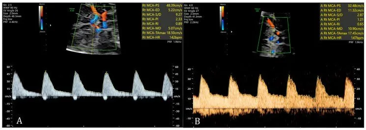
- Regardless of the PI, the peak systolic velocity (PSV) in the MCA is the crucial marker for fetal anaemia.
- A value of of ≥ 1.5 MoM, indicating cordocentesis suggests moderate to severe anaemia.
- Reasons for anaemia diagnostics include, for example, Rhesus incompatibility, a parvovirus B19 infection or TAPS in the anemic donor twin.
What it looks like
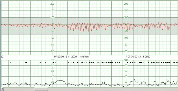
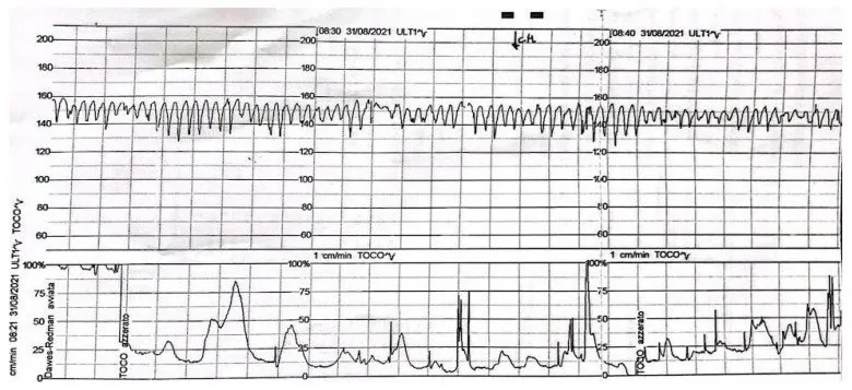
- The sinusoidal pattern is a smooth, sinusoidal line without variability.
- Amplitude and frequency are 5–15 bpm and 3–5 cycles/minute.
- The criterion is a duration of at least 30 minutes; accelerations are completely.
- Under FIGO the sinusoidal pattern counts straight away as pathological.
Typical imaging findings
- CTG: The trace runs in even, smooth waves around 130–140 beats, with a regular frequency of about three to five cycles per minute and constant amplitude. Short-term variability and accelerations are completely absent; the pattern persists over many minutes.
- CTG: Around a baseline of about 150 per minute runs an even, wave-like oscillation without any irregularity. Accelerations are absent and the tocogram shows only irregular small deflections.
- Non-invasively, the peak systolic velocity of the middle cerebral artery is measured; from 18 weeks a value above 1.5 MoM strongly suggests moderate to severe fetal anaemia.
- In alloimmunisation, antibody identification, the father's antigen status and serial titres are used for assessment; fetal haemoglobin is determined by umbilical cord blood sampling.
Keep learning in the app
Further reading (selection)
- StatPearls: Hemolytic Disease of the Fetus and Newborn (NCBI Bookshelf)
- MSD Manual Professional Edition: Hemolytic Disease of the Fetus and Neonate
- J Pers Med 2024: Parvovirus B19 Infection and Pregnancy – Review of the Current Knowledge (PMC, open access)
- DocCheck Flexikon, Arteria cerebri media
- DocCheck Flexikon, Kardiotokographie
Cross-references
More topics: Fetus & prenatal medicine
Note: Learning content from the GynFuchs app (flashcards, exam questions, image cases) for medical education – not a treatment recommendation and no substitute for diagnosis or treatment decisions in individual cases. Treatment and management are deliberately not covered on this page.