Omphalocele and gastroschisis
Exam relevance: in 10 of 197 board exam reports · rank 66
- Specialty
- Obstetrics · Fetus & prenatal medicine
- Images
- Ultrasound 5
- Exam relevance
- 10 of 197 reports · rank 66
- In the app
- 1 flashcards · GynFuchs
- Last updated
- 10/2026 · Dr. Pascal Bafteh
Contents
Images (5)
 Ultrasound
 Ultrasound
 Ultrasound
 Ultrasound
 Ultrasound
Definition
Characteristics of the defects
- An omphalocele is a midline defect in which the hernial sac is covered by amnion and peritoneum and the umbilical cord inserts on the sac.
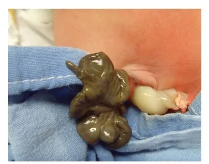
- Gastroschisis is typically a paraumbilical defect without a hernial sac, where intestinal loops float freely in the amniotic fluid.
- The small variant of an omphalocele is known as an umbilical cord hernia.
- Omphalocele is protrusion of abdominal viscera through a midline defect at the base of the umbilicus; the organs are covered by a thin membrane and range from a few bowel loops to stomach and liver.
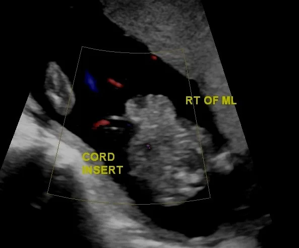
- Gastroschisis is a full-thickness paraumbilical abdominal wall defect, usually to the right of the cord insertion, with protrusion of bowel without a covering membrane.
Occurrence & epidemiology
Epidemiology
- Gastroschisis occurs in about 1 in 2,500 live births and is more common than omphalocele, whose incidence is about 2 to 3 per 10,000 live births.
Aetiopathogenesis
Aetiology and associated anomalies
- In omphalocele about two thirds have further anomalies, such as bowel atresia, chromosomal abnormalities (trisomy 18, 13 and 21), cardiac and renal anomalies or Beckwith-Wiedemann syndrome.
- Gastroschisis, by contrast, is rarely associated with genetic syndromes; only about 10% have anomalies outside the gastrointestinal tract, while gastrointestinal anomalies such as malrotation and intestinal atresia occur in up to a quarter.
Clinical features
Associations and further diagnostics
- In contrast to omphalocele, chromosomal anomalies in gastroschisis are rare.
Clinical features and complications
- In gastroschisis, direct exposure to amniotic fluid causes a chemical peritonitis with an oedematous, matted bowel wall and fibrinous rind, which may later lead to fibrosis, dysmotility or obstruction; it is considered complex in the presence of atresia, stenosis, perforation, necrosis, malrotation or volvulus.
- Infants with gastroschisis frequently have growth restriction.
Diagnosis
Differentiation in the first trimester
- A physiological midgut herniation is a normal finding until about the 11th to 12th week.
- A true abdominal wall defect does not resolve during the course of the pregnancy.
Typical imaging findings
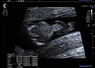
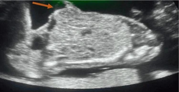
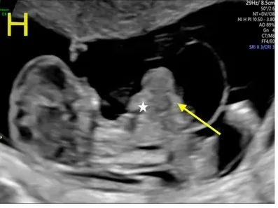
- Ultrasound: A midline sac bulges from the anterior abdominal wall, covered by a fine echogenic membrane. The umbilical cord inserts at the apex of the sac; the contents remain smoothly bordered and do not float freely.
- Ultrasound: A rounded, sharply outlined bulge with a smooth surface lies in front of the abdominal wall. The cord inserts centrally on this bulge and the contents are covered by a thin membrane.
- Ultrasound: To the right of the cord insertion bowel loops pass through the abdominal wall and lie free in the amniotic fluid without a covering membrane. Individual loops are distinguishable and float; the cord insertion itself is normal.
- Omphalocele is detected on routine prenatal ultrasound; gastroschisis typically on the ultrasound at about 20 weeks, showing bowel loops floating freely in the amniotic fluid.
- In gastroschisis, maternal serum alpha-fetoprotein is raised.
Keep learning in the app
Further reading (selection)
Cross-references
Note: Learning content from the GynFuchs app (flashcards, exam questions, image cases) for medical education – not a treatment recommendation and no substitute for diagnosis or treatment decisions in individual cases. Treatment and management are deliberately not covered on this page.