Fetal hypoxia and acidosis
Exam relevance: in 10 of 197 board exam reports · rank 66
- Specialty
- Obstetrics · Fetus & prenatal medicine
- Images
- CTG 8
- Exam relevance
- 10 of 197 reports · rank 66
- In the app
- 3 flashcards · GynFuchs
- Last updated
- 10/2026 · Dr. Pascal Bafteh
Contents
Images (8)
 CTG
CTG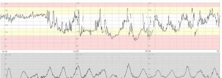 CTG
CTG CTG
CTG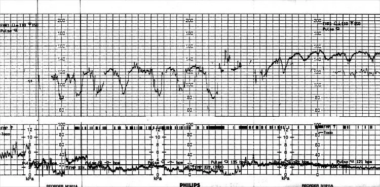 CTG
CTG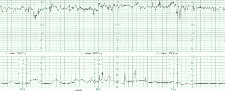 CTG
CTG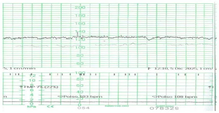 CTG
CTG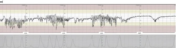 CTG
CTG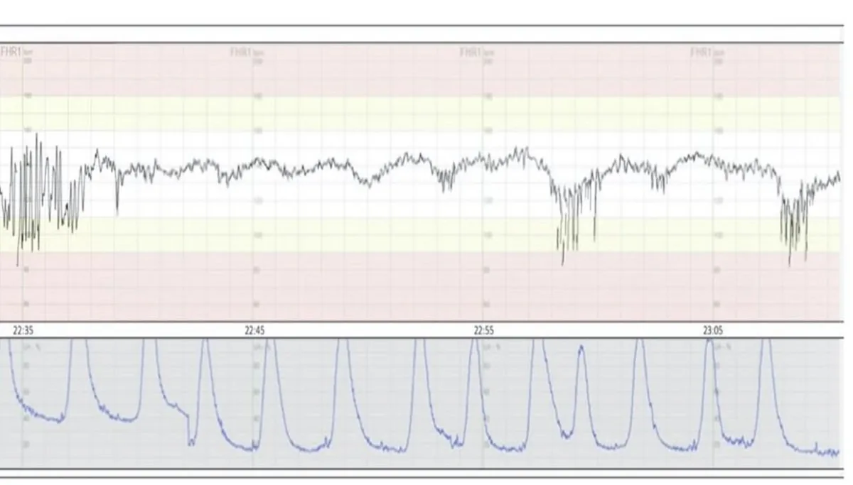 CTG
CTGDefinition
- Perinatal asphyxia is a lack of blood flow or gas exchange to or from the fetus immediately before, during or after birth.
- A partial lack of oxygen is called hypoxia and a complete lack anoxia; progressive hypoxaemia and hypercapnia follow.
Classification
FIGO CTG categories
- Normal means baseline 110–160, var. 5–25, no repet. decels.
- Suspicious means one feature of normality is missing.
- A CTG is pathological with baseline <100, var. <5/>25, sinusoid/repet. decels.
- "Repetitive" means the deceleration occurs with >50% of contractions.
Aetiopathogenesis
Aetiology and pathophysiology
- The cause is impaired blood flow or gas exchange across the placenta before birth or via the lungs after birth; the reduced supply affects the brain, heart, liver and muscles.
- A severe oxygen deficit triggers anaerobic metabolism with lactic acidosis; the neurological damage due to asphyxia and ischaemia is called hypoxic-ischaemic encephalopathy.
Clinical features
- A hypoxic event presents in the newborn with metabolic acidosis, base deficit, low Apgar scores and multi-organ failure.
- Signs of encephalopathy are hypotonia, abnormal oculomotor or pupillary responses, a weak or absent suck, apnoea, hyperpnoea and seizures; MRI shows characteristic findings.
Diagnosis
Decelerations
- A deceleration is a fall in heart rate of ≥15 bpm for ≥15 s.
- Early decelerations are shallow, uniform, mirroring the contraction and arise from head compression with vagal response.
- Variable decelerations change in shape, depth and timing and arise from umbilical cord compression.
- Late decelerations begin after the contraction's peak and suggest uteroplacental insufficiency.
Baseline
- The baseline is mean FHR over ≥10 min, without acc./dec..
- Normal is 110–160 bpm; with advancing gestation it it physiologically decreases.
- Fetal tachycardia above 160 bpm: causes include fever, infection, betamimetics, anemia.
Bradycardia
- A baseline below 110 bpm is suspicious; below 100 bpm it is pathological.
- Harmless causes are vagal response to head compression, low rate; the dangerous ones are abruption, rupture, cord prolapse and maternal hypotension.
Variability
- Normal oscillation bandwidth is at 5–25 bpm.
- A silent pattern below 5 bpm for more than 50 minutes is pathological.
- A saltatory pattern above 25 bpm for more than 30 minutes counts as a pathological finding.
Typical imaging findings
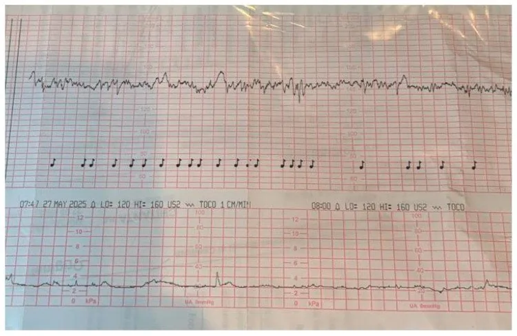
- CTG: The baseline lies at 125 to 130 per minute. After several contractions the rate dips with a delay to below 100 per minute and returns to baseline only once the contraction is over. The dips are shallow, uniform and recurrent.
- CTG: The baseline lies at about 160 per minute with preserved variability. After each contraction the heart rate falls with a delay; the nadir lies well behind the peak of the contraction and recovery drags on past the end of it.
- CTG: After an initially normal baseline the trace falls steeply and stays far below the starting level, without recovering within the minutes displayed. The fall is not tied to a single contraction and lasts considerably longer than a typical deceleration.
- CTG: After an initially normal stretch the heart rate drops steeply and stays well below the baseline for several minutes before recovering slowly. The fall is not tied to a single contraction and lasts longer than two minutes.
- On CTG the normal baseline is 110 to 160/min and normal (moderate) variability 6 to 25/min; a baseline above 160/min is tachycardia and below 110/min bradycardia.
- Category III is associated with fetal acidaemia: absent variability together with recurrent late or variable decelerations or bradycardia, and a sinusoidal pattern.
- Interpretation is highly observer-dependent; in one study obstetricians interpreted tracings consistently in only 29% of cases.
Keep learning in the app
Further reading (selection)
Cross-references
More topics: Fetus & prenatal medicine
Note: Learning content from the GynFuchs app (flashcards, exam questions, image cases) for medical education – not a treatment recommendation and no substitute for diagnosis or treatment decisions in individual cases. Treatment and management are deliberately not covered on this page.