Fetal renal anomalies
Exam relevance: in 2 of 197 board exam reports · rank 144
- Specialty
- Obstetrics · Fetus & prenatal medicine
- Images
- Ultrasound 1 · Clinical 1
- Exam relevance
- 2 of 197 reports · rank 144
- Last updated
- 10/2026 · Dr. Pascal Bafteh
Contents
Images (2)
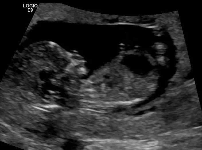 Ultrasound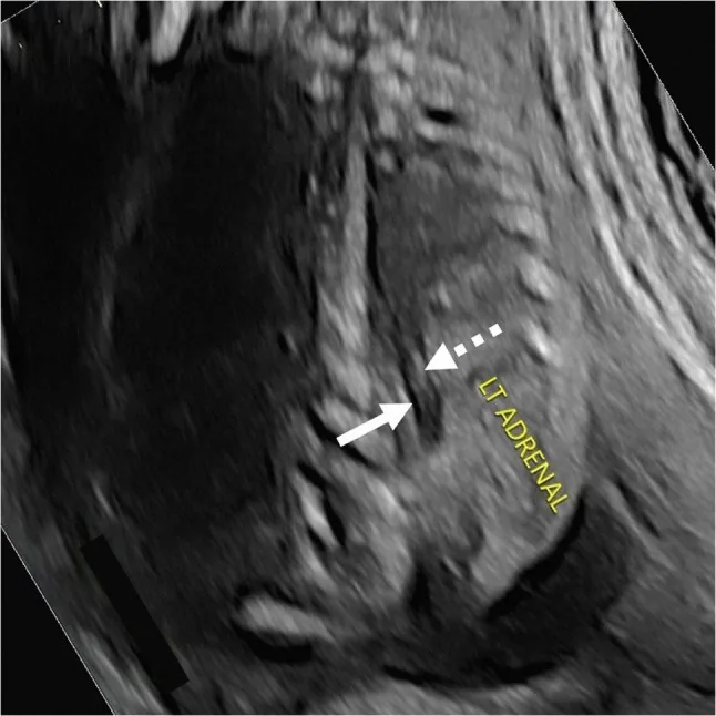
Definition
- Congenital anomalies of the kidneys and urinary tract range from renal agenesis through cystic kidney disease to urinary tract obstruction.
- Potter sequence describes the consequences of reduced amniotic fluid regardless of the cause; in Potter syndrome the oligohydramnios results from renal agenesis or impaired renal function.
Classification
- Bilateral renal agenesis is classic for Potter syndrome; type I is associated with autosomal recessive polycystic kidney disease, type II with renal dysplasia, type III with autosomal dominant polycystic kidney disease and type IV with obstruction of the ureter or renal pelvis with hydronephrosis.
Occurrence & epidemiology
Epidemiology
- Posterior urethral valves occur exclusively in males and are the most common cause of urinary tract obstruction and of chronic kidney disease due to obstructive uropathy in childhood.
Aetiopathogenesis
Aetiology and pathophysiology
- Because fetal urine contributes to amniotic fluid volume, absent urine production or severe urethral obstruction leads to oligohydramnios, which causes pulmonary hypoplasia with pulmonary hypertension.
- Posterior urethral valves are obstructing membranous folds in the posterior urethra attached to the verumontanum; the commonest type I (95%) is regarded as a remnant of the Wolffian duct.
Clinical features
Clinical features and complications
- Potter syndrome is incompatible with life: pulmonary hypoplasia causes respiratory distress within an hour of birth; typical facial and limb changes are also present.
- Posterior urethral valves range from asymptomatic to non-viable forms; complications include urinary retention, hydroureteronephrosis, vesicoureteral reflux, renal failure and, in severe cases, pulmonary hypoplasia up to perinatal death.
Diagnosis
- Posterior urethral valves are often suspected on routine prenatal ultrasound, for example with severe bilateral hydroureteronephrosis or oligohydramnios; after birth the diagnosis is confirmed by voiding cystourethrography.
Keep learning in the app
Further reading (selection)
Cross-references
Note: Learning content from the GynFuchs app (flashcards, exam questions, image cases) for medical education – not a treatment recommendation and no substitute for diagnosis or treatment decisions in individual cases. Treatment and management are deliberately not covered on this page.