Cleft lip and palate (prenatal)
- Specialty
- Obstetrics · Fetus & prenatal medicine
- Images
- Ultrasound 1
- Last updated
- 10/2026 · Dr. Pascal Bafteh
Contents
Images (1)
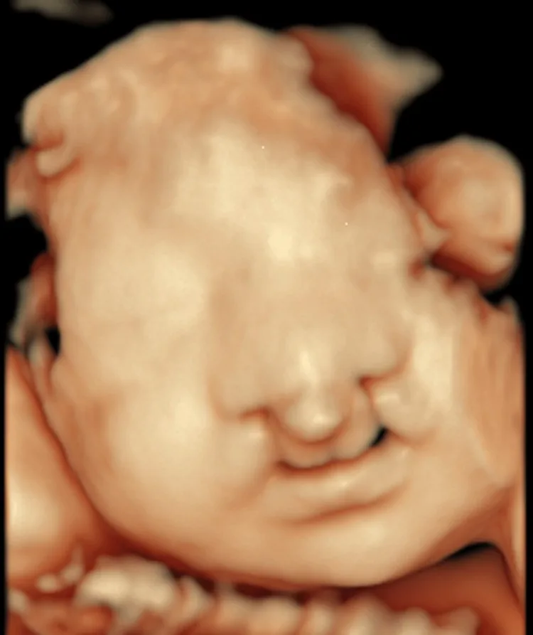
Definition
- The clefts develop during weeks 6 to 8; the primary and secondary palate have different embryological origins.
Classification
- Cleft lip with or without cleft palate is distinguished from isolated cleft palate; the spectrum ranges from bifid uvula as the mildest form through clefts of the soft palate only to complete clefts of the soft and hard palate, alveolar process and lip.
- About 30% of clefts are syndromic and about 70% non-syndromic (isolated).
Occurrence & epidemiology
Epidemiology
- Together, cleft lip and palate are the most common congenital anomalies of the head and neck, with a total prevalence of about 1 in 600 to 1,000 live births; they are most frequent in people of Asian and Native American origin.
Aetiopathogenesis
Aetiology and risk factors
- Genetic and environmental factors are involved; syndromic clefts are typically caused by chromosomal abnormalities or defined monogenic syndromes.
- Risk factors include smoking and alcohol use in pregnancy, a cleft in the mother or a first-degree relative and possibly folate deficiency, including from folate antagonists such as phenytoin, valproate or carbamazepine.
Clinical features
- Clefts impair feeding through nasal reflux, an inadequate latch and increased work of feeding with fatigue; later, speech development is impaired and middle ear infections are more frequent.
Diagnosis
- Prenatally, clefts are sought with 2D ultrasound; supplementary 3D ultrasound or MRI improves diagnostic accuracy in most studies.
- After birth, besides clinical examination, genetic evaluation is undertaken even for apparently isolated clefts, for example with chromosomal microarray analysis or gene panels.
Keep learning in the app
Further reading (selection)
Cross-references
Note: Learning content from the GynFuchs app (flashcards, exam questions, image cases) for medical education – not a treatment recommendation and no substitute for diagnosis or treatment decisions in individual cases. Treatment and management are deliberately not covered on this page.