Deep vein thrombosis
Overview
Clinical Signs
- Payr: Pain with flexion / pressure on the medial sole of the foot
- Meyer (= Mayr): Pain with pressure on the calf
- Homans: Pain medial sole of the foot / calf with dorsiflexion
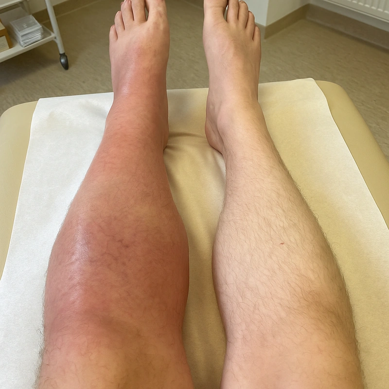
- Local: warm, edematous, livid leg, prominent collateral veins
Wells Score (DVT)
| Point | Criterion |
|---|---|
| +1 | active cancer |
| +1 | paralysis / recent immobilization |
| +1 | bed rest > 3 d / major surgery < 12 wks (per original Wells, Braun-Falco p. 1221) |
| +1 | pain along venous distribution |
| +1 | entire leg swelling |
| +1 | calf circumference ≥ 3 cm difference |
| +1 | pitting edema more pronounced in symptomatic leg |
| +1 | collateral veins visible |
| +1 | previous objectively confirmed DVT |
| −2 | alternative diagnosis at least as likely |
- Assessment: ≥ 2 → DVT likely → Duplex ultrasound ± D-dimer. If < 2 + neg. D-dimer → DVT unlikely.
- Surgery time window (common pitfall!): In the original Wells score, major surgery < 12 weeks counts as +1 point — not "< 4 weeks", as is often incorrectly taught.
D-dimer — for exclusion only, never for proof
- The D-dimer test has high sensitivity and low specificity. It is therefore useful only to exclude DVT when pre-test probability is low — a positive value proves nothing, a negative value with Wells < 2 practically rules DVT out.
- Age-adjusted cut-off from 50 years on: instead of the fixed 500 µg/l FEU cut-off, use age × 10 µg/l — so 800 µg/l in an 80-year-old. This markedly reduces unnecessary ultrasound scans without missing thromboses.
- False positives occur in pregnancy, cancer, inflammation, recent surgery, trauma and old age — that is, in nearly every patient in whom it gets ordered.
Compression Ultrasonography (CUS) — the standard test for DVT
- Principle: A patent vein can be fully compressed with gentle probe pressure in the transverse plane (the lumen disappears and only the pulsating artery remains); a thrombosed vein is not or only incompletely compressible — non-compressibility is the decisive criterion, not echogenicity.
- Procedure: in the transverse plane from proximal to distal in 1–2 cm steps: common femoral vein including the saphenofemoral junction (crosse) → femoral vein → adductor canal → popliteal vein (patient seated or prone) → calf veins (posterior tibial and fibular veins). Colour duplex and flow manoeuvres (Valsalva, distal compression) are added to assess residual lumen and reflux. Whole-leg compression ultrasound covers the entire limb, whereas two-point compression ultrasound examines only groin and popliteal fossa — with a negative result and persisting suspicion a repeat scan after 5–7 days is required.
- Vein wall structure: three layers as in the artery — tunica intima, tunica media, tunica adventitia. Compared with the artery the media is far less muscular and much thinner — which is why a vein is compressible at all; from medium calibre upwards veins additionally carry valves (intimal duplications).
- Thrombus age from echogenicity: A fresh thrombus is hypoechoic to almost anechoic and is therefore easily missed — it betrays itself only by the loss of compressibility and the distended vein lumen. With increasing age the thrombus becomes more echogenic, shrinks and adheres to the wall; a mural, echogenic residue indicates a post-thrombotic residual finding rather than fresh thrombosis.
Differential diagnoses
- Great saphenous vein varicosity
- Telangiectasias (spider veins) and reticular veins
- Corona phlebectatica paraplantaris
- Purpura jaune d’ocre
- Atrophie blanche
- Lipodermatosclerosis
Practise Deep vein thrombosis in the app
Flashcards with spaced repetition, exam questions and spot-the-diagnosis on this topic – in DermaFuchs, free of charge.
In the DermaFuchs app: 1 flashcards · 2 clinical images
Open in browser Download on the App StoreSources (selection)
- Wells 2003 NEJM D-Dimer DVT
- Plewig 2018 Braun-Falco p.1221
- Linnemann B, et al. Diagnostik und Therapie der Venenthrombose und der Lungenembolie. S2k-Leitlinie, AWMF-Register-Nr. 065/002. Stand: September 2023.
More vascular skin disorders
- Erythromelalgia
- Leukocytoclastic vasculitis
- Lichen aureus
- Superficial thrombophlebitis
- Great saphenous vein varicosity
Note: This page is intended for medical education and does not replace diagnosis or treatment decisions in individual cases. Treatment and follow-up content is available in the app.