Silicosis
- Synonyms
- dust lung, quartz dust lung, stonemason's lung, pneumoconiosis, silica lung disease
- Specialty
- Internal medicine · Pulmonology
- Images
- X-ray 2 · Histology 1 · Gross specimen 1
- Last updated
- 10/2026 · Dr. Pascal Bafteh
Contents
Images (4)
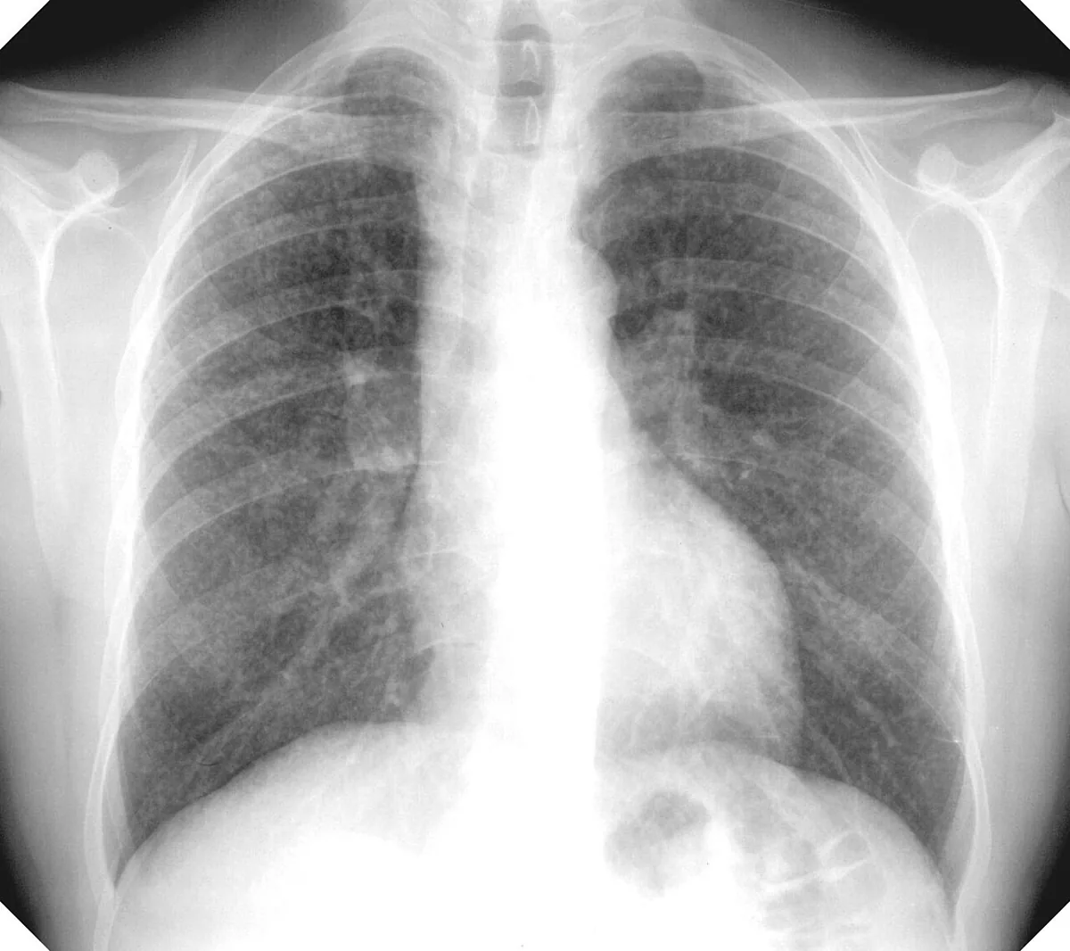 X-ray
X-ray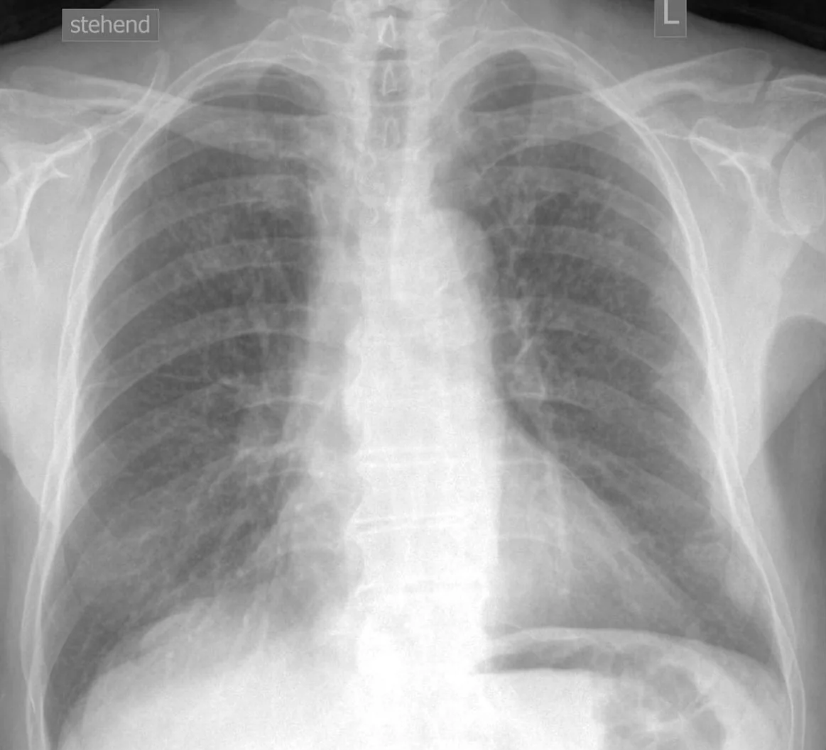 X-ray
X-ray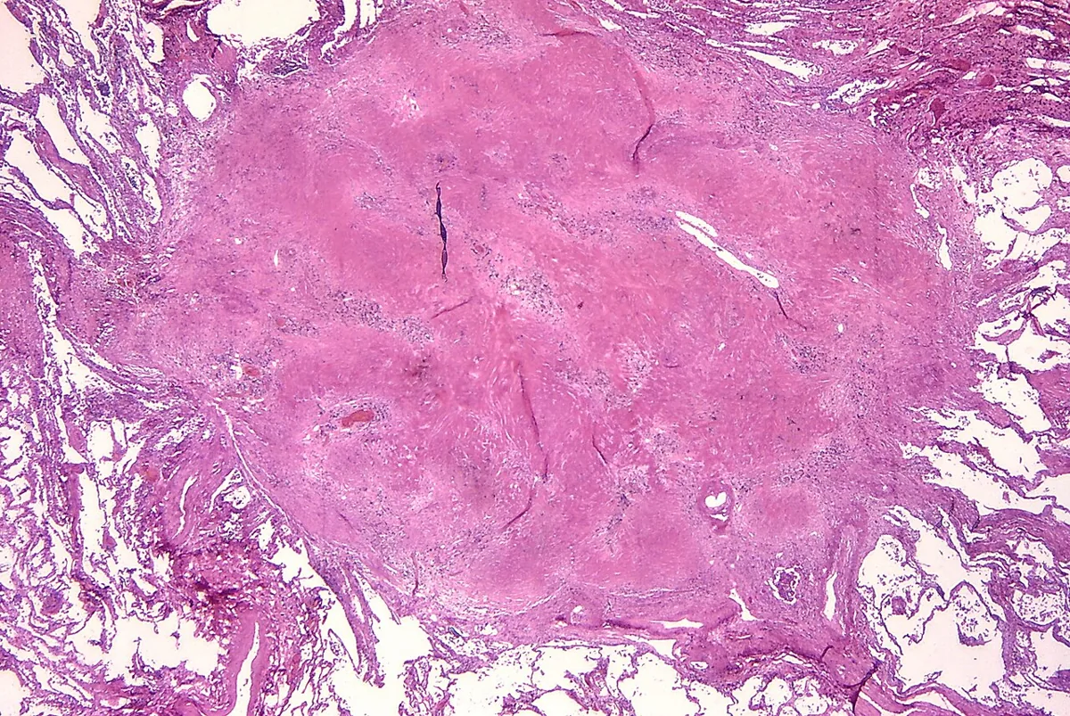 Histology
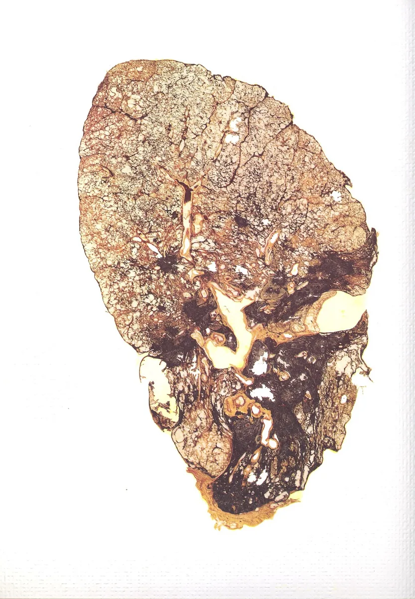 Gross specimen
Definition
Silicosis is a pneumoconiosis caused by inhaling respirable crystalline silicon dioxide (mostly quartz). It is characterized by nodular pulmonary fibrosis. Amorphous silica such as glass or diatomaceous earth has no crystalline structure and does not cause silicosis.
Classification
- Chronic silicosis: the most common form, develops slowly and typically appears decades after first exposure. In the simple form the nodules remain discrete and do not impair lung function; in the complicated form (progressive massive fibrosis) they coalesce into large conglomerates, usually in the upper zones.
- Accelerated silicosis: like the chronic form but with high exposure and onset within 5–10 years of first exposure.
- Acute silicosis (silicoproteinosis): after intense exposure over a short time; onset weeks to a few years after first exposure, resembling pulmonary alveolar proteinosis.
Aetiopathogenesis
Most at risk are people who move or blast rock and sand (miners, quarry workers, stonemasons, construction workers) or use silica-containing abrasives (sandblasters, glassmakers, foundry workers, gemstone and ceramic workers, potters). Severe outbreaks have also been observed in the natural stone industry. Incidence and severity depend on the duration and intensity of exposure and on the shape and surface properties of the particles.
Pathophysiology: Alveolar macrophages take up the silica particles and release cytokines, reactive oxygen species and other mediators that promote inflammation and fibrosis. Silica-laden macrophages promote the formation of the pathognomonic silicotic nodule.
Clinical features
- Chronic silicosis: may be asymptomatic; many patients develop progressive breathlessness and cough. In advanced disease consolidation, pulmonary hypertension and respiratory failure with or without right heart failure; in progressive massive fibrosis severe chronic respiratory symptoms.
- Acute silicosis: rapidly increasing breathlessness, loss of weight and exhaustion with diffuse bilateral crackles; hypoxemia is common.
- Complications: increased risk of tuberculosis and other mycobacterial infections, COPD, lung cancer and systemic autoimmune diseases.
Histology
The silicotic nodule initially contains collagen fibers and scattered birefringent silica particles, best seen with polarized light microscopy. As it matures, the collagen fibers lie centrally and are surrounded by an outer layer of inflammatory cells.
Diagnosis
- Occupational history: duration of exposure, protective measures at work and time course of symptoms; silicosis is still underdiagnosed.
- Chest X-ray: graded according to the ILO classification of pneumoconioses. In simple silicosis predominantly upper-lobe, bilateral, 1–3 mm reticulonodular opacities; calcified hilar and mediastinal lymph nodes, occasionally eggshell-like (not pathognomonic). In complicated silicosis large opacities or calcified conglomerates.
- Chest CT: more sensitive than the X-ray; in acute silicosis diffuse alveolar, basal-predominant opacities resembling alveolar proteinosis.
Keep learning in the app
Further reading (open access)
Cross-references
Note: Learning content for medical education – not a treatment recommendation and no substitute for diagnosis or treatment decisions in individual cases. Treatment and management are deliberately not covered on this page.