Lung abscess
- Synonyms
- pulmonary abscess, lung cavity infection, aspiration abscess, pulmonary gangrene
- Specialty
- Internal medicine · Pulmonology
- Images
- Diagram 1 · CT 1 · X-ray 1 · Gross specimen 1
- Last updated
- 10/2026 · Dr. Pascal Bafteh
Contents
Images (4)
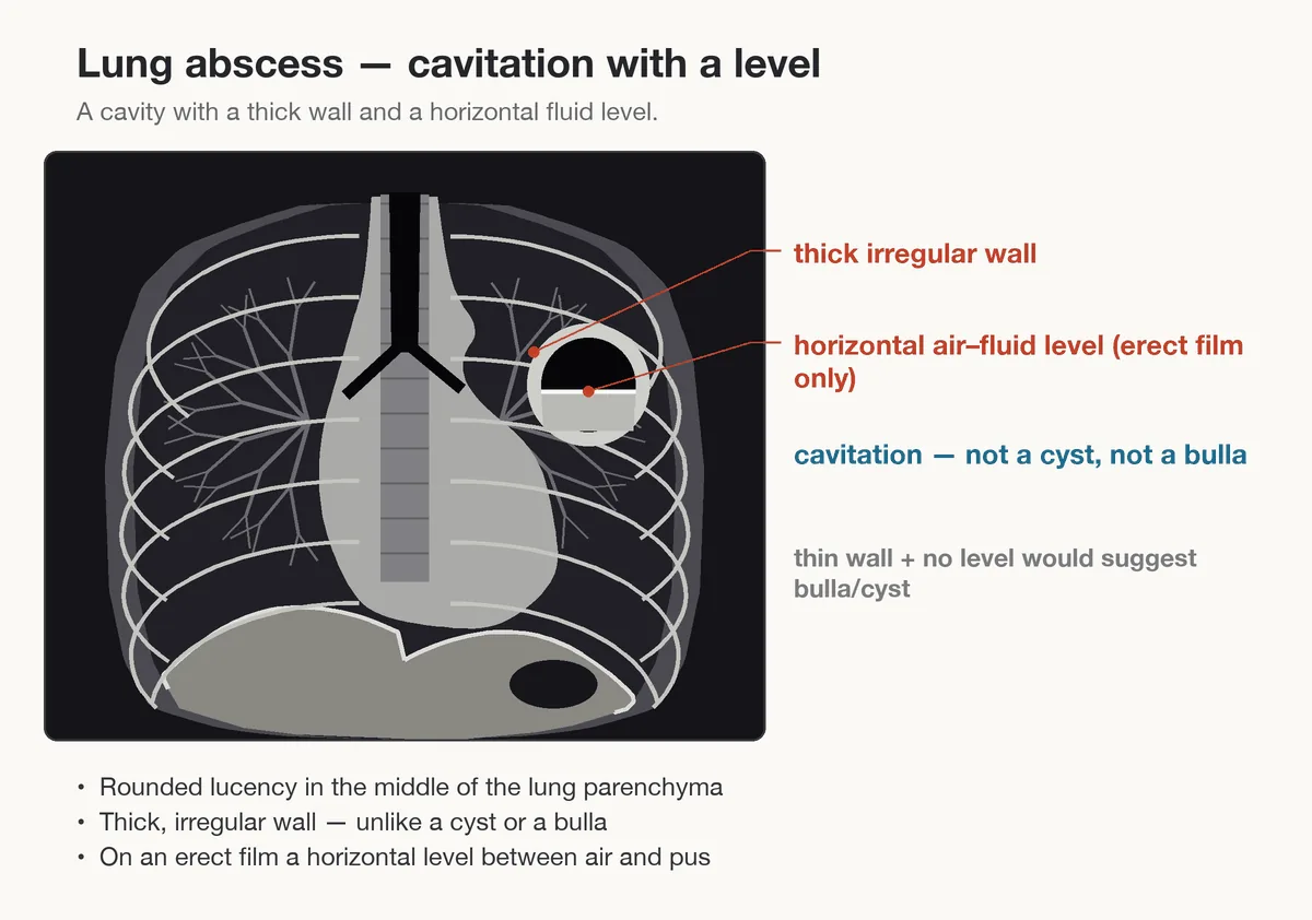 Diagram
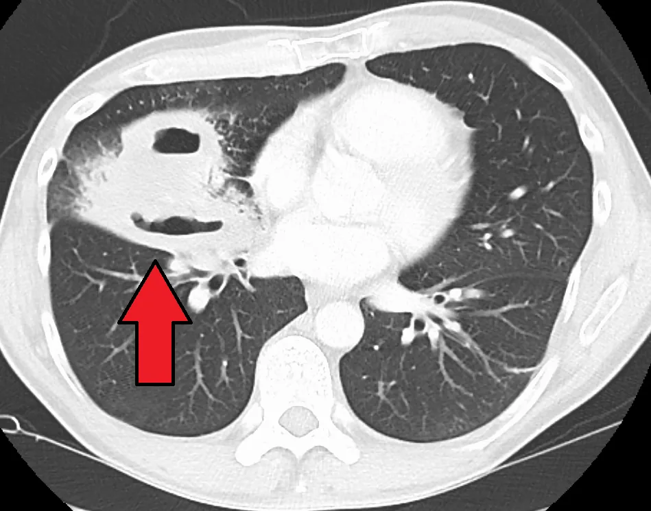 CT
CT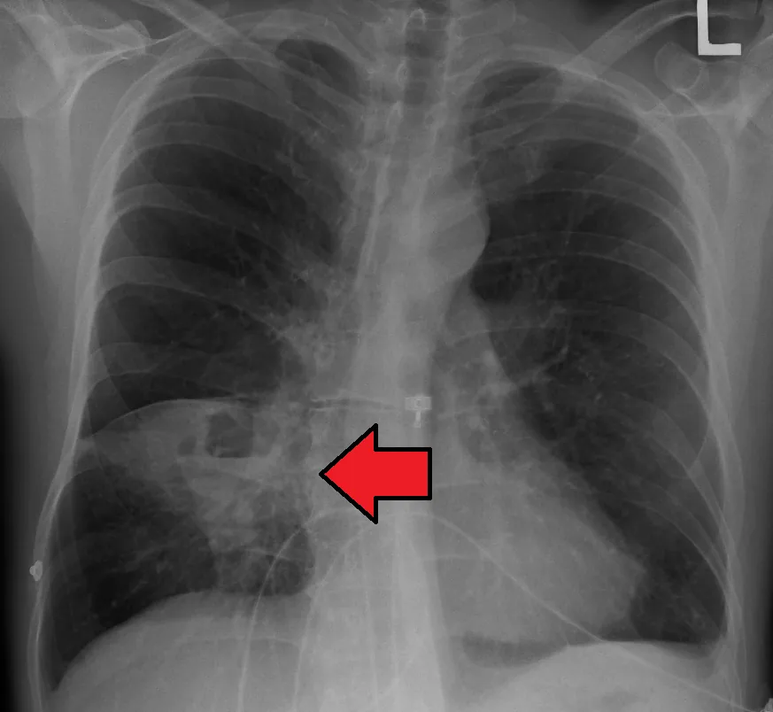 X-ray
X-ray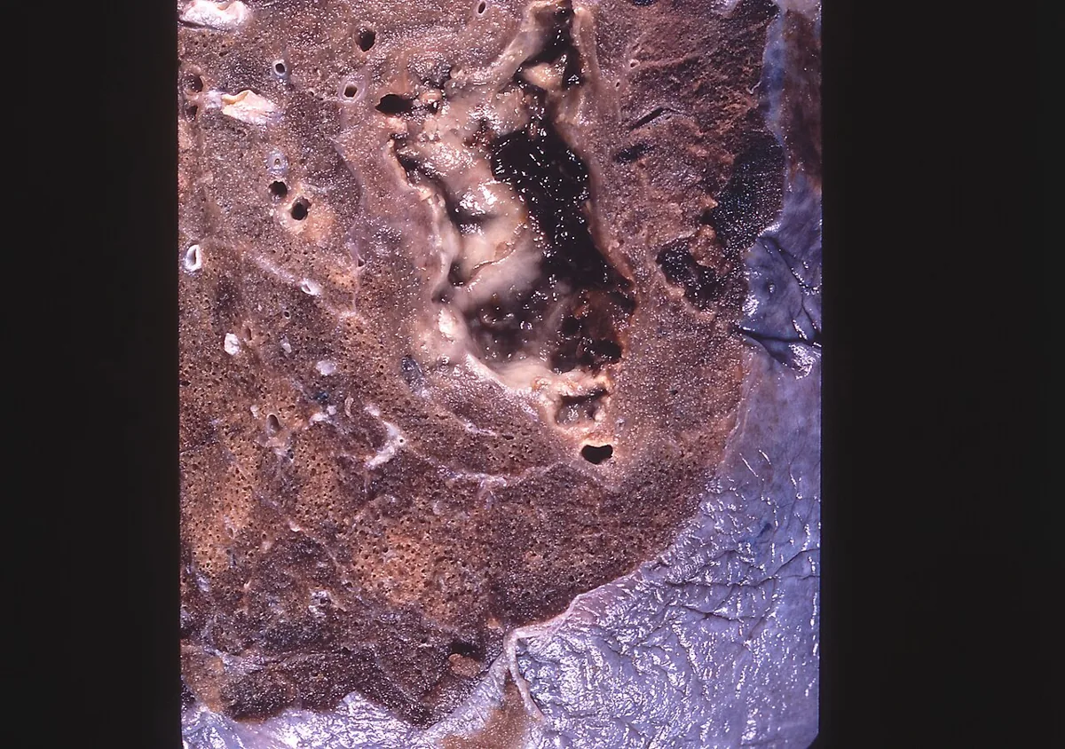 Gross specimen
Definition
A lung abscess is a necrotizing lung infection with a circumscribed, pus-filled cavity. It usually develops after aspiration of oral secretions in people prone to aspiration and has an insidious onset. Abscesses may arise from necrotizing pneumonia or pulmonary gangrene.
Aetiopathogenesis
- Aspiration of oral secretions (most common cause): with gingivitis or poor oral hygiene, usually together with impaired consciousness from alcohol, drugs, anesthesia, sedatives or opioids; also in older people and those with swallowing disorders, often due to neurological disease.
- Endobronchial obstruction: e.g. by bronchial carcinoma or foreign body.
- Immunosuppression: e.g. in advanced HIV infection or after organ grafting.
- Hematogenous seeding (less common): septic emboli, e.g. with drug use, Lemierre syndrome or right-sided endocarditis; usually acute and with multiple abscesses.
- Direct spread: from an empyema, a subphrenic or mediastinal abscess.
Pathogens: With aspiration, anaerobes predominate (Peptostreptococcus, Fusobacterium, Prevotella, Bacteroides); in about half of cases anaerobes and aerobes are involved together. Common aerobic pathogens are streptococci and staphylococci (sometimes MRSA), occasionally gram-negative bacteria such as Klebsiella. In immunocompromised patients Pseudomonas aeruginosa and other gram-negative pathogens, Nocardia, mycobacteria and fungi are added; rarely amoebae, Echinococcus or Paragonimus.
Pathogenesis: The inflammatory reaction leads over one to two weeks to tissue necrosis and abscess formation. Usually the abscess drains into a bronchus, leaving a cavity filled with air and fluid.
Clinical features
- Anaerobic or mixed: usually chronic over weeks to months with productive cough, fever, chills, night sweats and loss of weight; purulent or blood-tinged sputum that smells and tastes foul, halitosis; hemoptysis, pleuritic pain, loss of appetite and fatigue; digital clubbing possible.
- Aerobic pathogens: a more acute course over hours to days, similar to bacterial pneumonia.
- Mycobacteria or Nocardia: no foul secretions, subacute course, more often in non-dependent lung regions.
- Findings: nonspecific as in pneumonia – signs of consolidation, temperature ≥ 38 °C, crackles; usually signs of periodontal disease and a history of aspiration risk.
- Complications: empyema in about 10 % of cases through direct extension or a bronchopleural fistula.
Diagnosis
- Chest X-ray: with aspiration, typically consolidation with a single cavity and an air-fluid level in the lung regions that are dependent when lying down (posterior segment of the upper lobe, superior or laterobasal segment of the lower lobe). Embolic processes tend to cause multiple cavities.
- Chest CT: when a cavity is not clear on X-ray, and when an obstructing mass is suspected.
- Microbiology: Gram stain and culture of sputum and blood, cultures for fungi and mycobacteria if clinically suspected; with effusion, Gram stain and culture of the pleural fluid.
- Bronchoscopy: to exclude carcinoma or a foreign body, to detect unusual pathogens and in immunocompromised patients.
- Echocardiography: with multiple abscesses, to look for infective endocarditis.
Keep learning in the app
Further reading (open access)
Cross-references
Note: Learning content for medical education – not a treatment recommendation and no substitute for diagnosis or treatment decisions in individual cases. Treatment and management are deliberately not covered on this page.