Hospital-acquired pneumonia (HAP/VAP)
Board exam relevance: in 1 of 105 exam reports · rank 181- Synonyms
- HAP, VAP, ventilator-associated pneumonia, nosocomial pneumonia, hospital pneumonia
- Specialty
- Internal medicine · Pulmonology
- Images
- Gross specimen 2
- Last updated
- 10/2026 · Dr. Pascal Bafteh
Contents
Images (2)
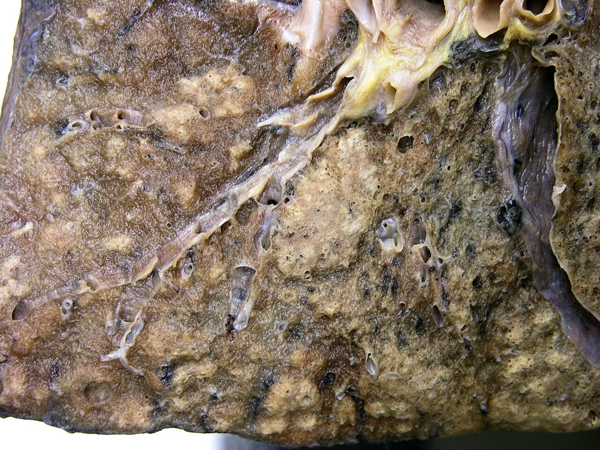 Gross specimen
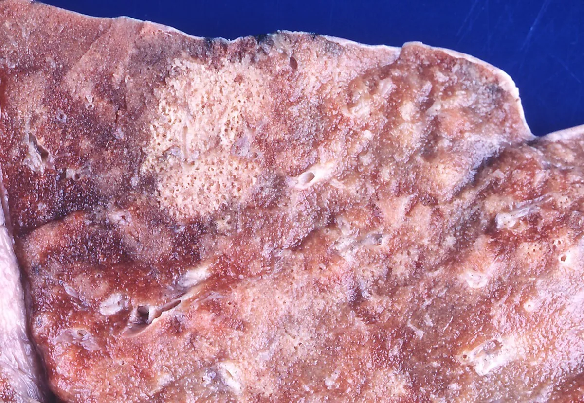 Gross specimen
Definition
Hospital-acquired (nosocomial) pneumonia (HAP) develops at least 48 hours after being admitted to hospital and was not incubating at the time of hospital entry. Postoperative pneumonia is included.
VAP is a subset of hospital-acquired pneumonia: it arises at least 48 hours after placement of an endotracheal tube or within 48 hours of extubation. Organizms with reduced susceptibility to anti-infective agents occur more often in VAP.
Occurrence & epidemiology
VAP occurs in 9–27 % of patients receiving mechanical respiratory support via an endotracheal tube. The risk is highest in the first ten days after placement of the tube.
Aetiopathogenesis
Development: The most common cause is microaspiration of bacteria that colonize the oropharynx, upper airways and partly the upper gastrointestinal tract of severely ill patients. Less often, pathogens reach the lung via bacteremia or contaminated aerosols (e.g. containing legionellae, Aspergillus or influenza viruses). An endotracheal tube breaches airway defences, impairs coughing and mucociliary clearance and facilitates aspiration of secretions above the tube cuff; bacteria form a protective biofilm on the tube.
Pathogens: mainly gram-negative bacilli (Pseudomonas aeruginosa, Enterobacter, Klebsiella pneumoniae, Escherichia coli, Serratia, Proteus, Acinetobacter) and Staphylococcus aureus including MRSA. In pneumonia within 4–7 days of admittance, methicillin-sensitive S. aureus, Streptococcus pneumoniae and Hemophilus influenzae predominate; with longer stay, P. aeruginosa, MRSA and enteric gram-negative bacteria become more frequent. Anaerobes play no major role. The spectrum of pathogens differs considerably between institutions.
Risk factors: older age, impaired consciousness, aspiration, previous anti-infective agents, raised gastric pH, heart, lung, liver or kidney failure; for postoperative pneumonia age over 70 and an upper abdominal or thoracic wound site. Chronic suppurative lung diseases such as cystic fibrosis and bronchiectasis favor gram-negative pathogens.
Clues to multidrug-resistant organizms in VAP: parenteral anti-infective exposure within the past 90 days, septic shock at the time of VAP, preceding ARDS and a hospital stay of at least 5 days before VAP develops.
Clinical features
- Without an endotracheal tube similar to community-acquired pneumonia: malaise, fever, rigors, cough, breathlessness and chest pain
- With an endotracheal tube: fever, rising respiratory or heart rate, increasing purulent secretions, worsening hypoxemia
Diagnosis
The diagnosis is usually based on a new lung infiltrate on chest X-ray or CT together with clinical signs of infection: recurrent fever, purulent sputum, leukocytosis and worsening oxygenation. No symptom or X-ray finding alone is sufficiently sensitive or specific.
- Sputum or tracheal aspirate: Gram stain and semiquantitative culture.
- Bronchoscopically obtained samples from the lower airways with quantitative culture: distinguish more reliably between colonization and infection.
- Blood cultures: relatively specific when a respiratory pathogen grows, but insensitive.
- Pleural fluid: with a pleural effusion, culture of a pathogen from the pleural fluid is the only method that reliably confirms both pneumonia and pathogen.
- Molecular tests: detect pathogens and genetic markers of reduced susceptibility in respiratory secretions.
Keep learning in the app
Further reading (open access)
Cross-references
Note: Learning content for medical education – not a treatment recommendation and no substitute for diagnosis or treatment decisions in individual cases. Treatment and management are deliberately not covered on this page.