Bronchiectasis
- Synonyms
- bronchiectases, dilated airways, non-CF bronchiectasis, widened bronchi
- Specialty
- Internal medicine · Pulmonology
- Images
- Clinical 1 · CT 1 · Gross specimen 1
- Last updated
- 10/2026 · Dr. Pascal Bafteh
Contents
Images (3)
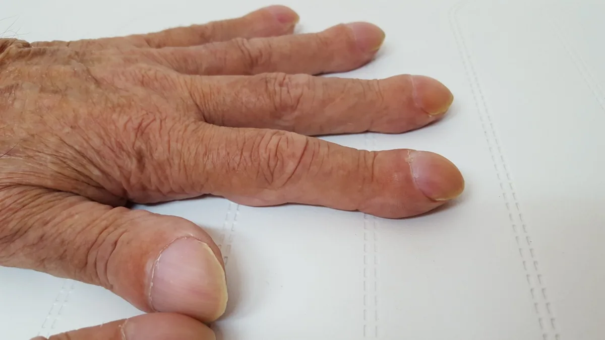
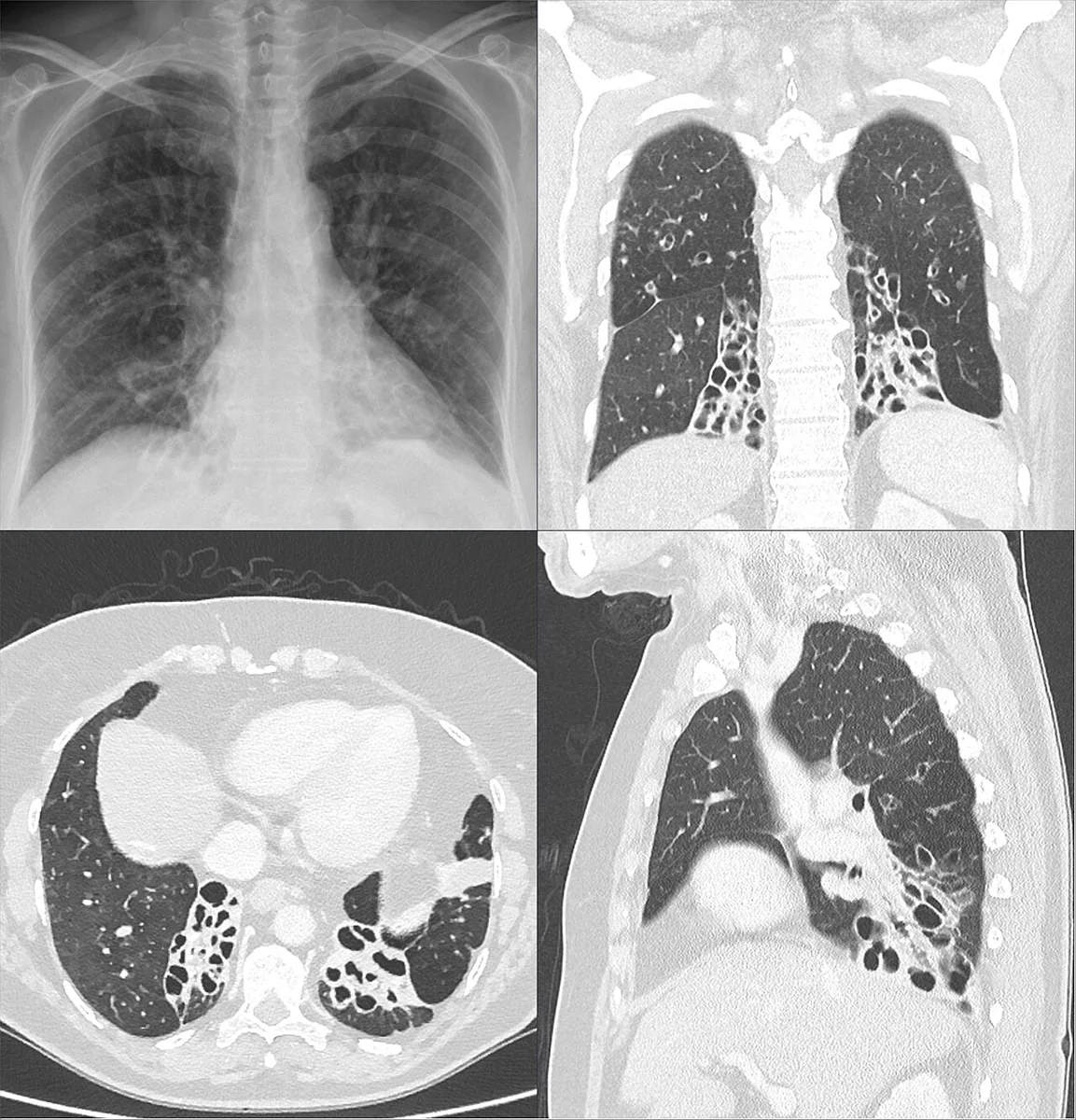 CT
CT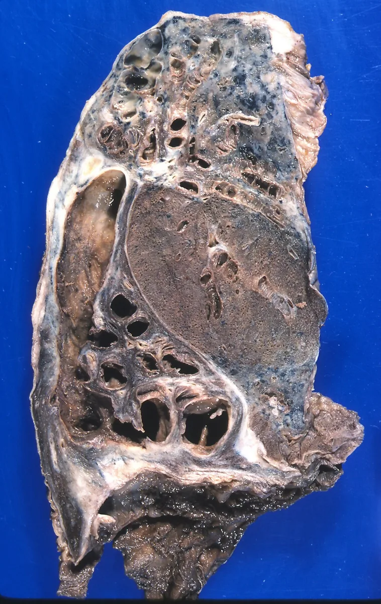 Gross specimen
Definition
Bronchiectasis is irreversible dilation and damage of the bronchi. It is regarded as the common end point of various diseases that cause chronic airway inflammation and may be diffuse (many lung regions) or focal (one or two regions). Traction bronchiectasis in pulmonary fibrosis is a secondary finding and is not counted as primary bronchiectasis.
Occurrence & epidemiology
Worldwide, an estimated 680 per 100,000 people are affected by bronchiectasis.
Aetiopathogenesis
- Infections: bacterial (e.g. Hemophilus influenzae, Pseudomonas aeruginosa, Staphylococcus aureus, Bordetella pertussis), mycobacterial (tuberculosis, non-tuberculous mycobacteria), viral (e.g. measles, adenoviruses, influenza) and fungal.
- Airway obstruction: endobronchial tumor, external compression by tumor or lymph nodes, foreign body; usually focal.
- Congenital disorders: cystic fibrosis (up to 20 % of initially idiopathic cases), primary ciliary dyskinesia (Kartagener syndrome with situs inversus), alpha-1 antitrypsin deficiency, Marfan syndrome.
- Immune defects: common variable immunodeficiency (CVID) and other hypogammaglobulinemias, HIV infection, hematological malignancies.
- Systemic diseases: rheumatoid arthritis, Sjögren syndrome, systemic lupus erythematosus, inflammatory bowel disease.
- Others: allergic bronchopulmonary aspergillosis (ABPA), chronic aspiration and gastresophageal reflux, advanced COPD or asthma, inhalation of toxic gases.
Pathophysiology: According to the "vicious cycle" model, an initial insult leads to inflammation and epithelial damage; the structural changes impair mucociliary clearance and promote bacterial colonization and infection. Proteases, especially elastase from neutrophils, destroy elastin, cartilage and muscle of the larger airways and lead to irreversible dilation.
Clinical features
- Insidious onset with gradual worsening over years; a small proportion is asymptomatic
- Chronic cough with thick, viscous, often purulent sputum as the most common symptom; breathlessness, wheeze, pleuritic pain
- Hemoptysis from neovascularization of the airways, sometimes massive
- Exacerbations: increased cough, breathlessness, sputum volume and purulence, often with low-grade fever and general symptoms
- Findings: halitosis, wheezes and rhonchi, dullness over obstructed areas; digital clubbing rare (more often in cystic fibrosis); chronic rhinosinusitis and nasal polyps in cystic fibrosis or primary ciliary dyskinesia
- Late stage: hypoxemia, pulmonary hypertension and right heart failure
Diagnosis
- Chest X-ray: thickened airway walls and dilated airways, indistinct ring shadows, "tram lines" and mucus-filled tubular opacities; upper-lobe predominant in cystic fibrosis.
- HRCT: the preferred method for confirmation and extent; airway diameter larger than the accompanying artery (signet ring sign), bronchi that do not narrow towards the periphery and reach almost to the pleura, tram lines; progression from cylindrical to varicose to cystic forms.
- Lung function: may be normal; the most common finding is obstruction.
- Sputum culture: for bacteria and mycobacteria.
Search for the cause
- Cystic Fibrosis (CF): In adults often atypical courses, sweat test and CFTR gene analysis.
- Primary Ciliary Dyskinesia (PCD): Kartagener syndrome (situs inversus, sinusitis, bronchiectasis), nasal NO measurement.
- Immunodeficiencies: Especially hypogammaglobulinemia, IgG subclass determination.
- Allergic bronchopulmonary aspergillosis (ABPA): In asthma and CF, elevated IgE levels, eosinophilia, Aspergillus precipitins.
- Post-infectious: After severe pneumonia (e.g., mycobacteria), pertussis.
- Further: Alpha-1 antitrypsin deficiency, rheumatoid arthritis, inflammatory bowel diseases.
Keep learning in the app
Further reading (open access)
Cross-references
Note: Learning content for medical education – not a treatment recommendation and no substitute for diagnosis or treatment decisions in individual cases. Treatment and management are deliberately not covered on this page.