Asbestosis and pleural mesothelioma
Board exam relevance: in 1 of 105 exam reports · rank 181- Synonyms
- asbestos lung, pleural mesothelioma, mesothelioma, pleural plaques, asbestos fibrosis
- Specialty
- Internal medicine · Pulmonology
- Images
- Gross specimen 1 · Histology 2 · CT 1
- Last updated
- 10/2026 · Dr. Pascal Bafteh
Contents
Images (4)
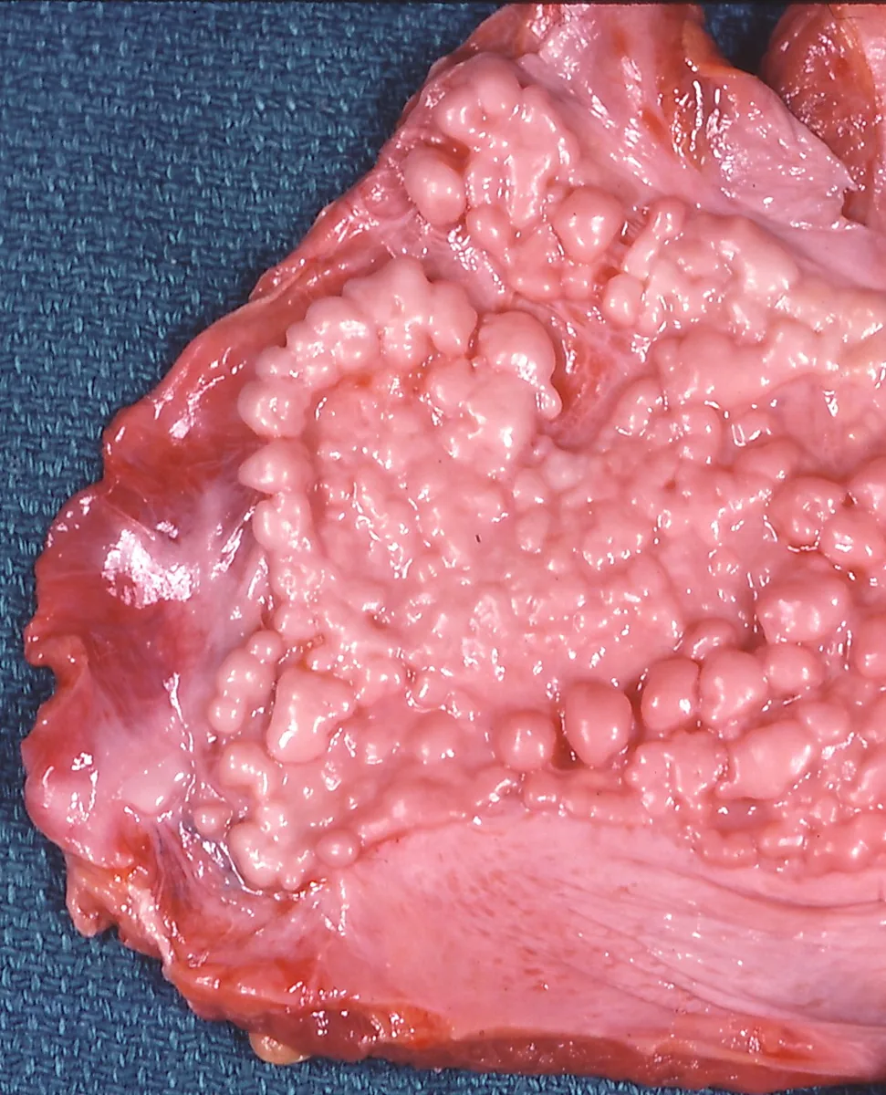 Gross specimen
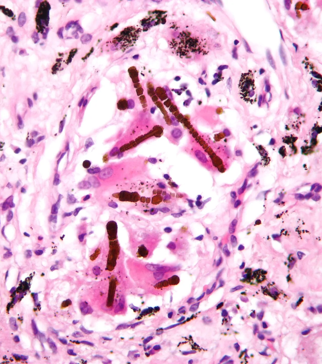 Histology
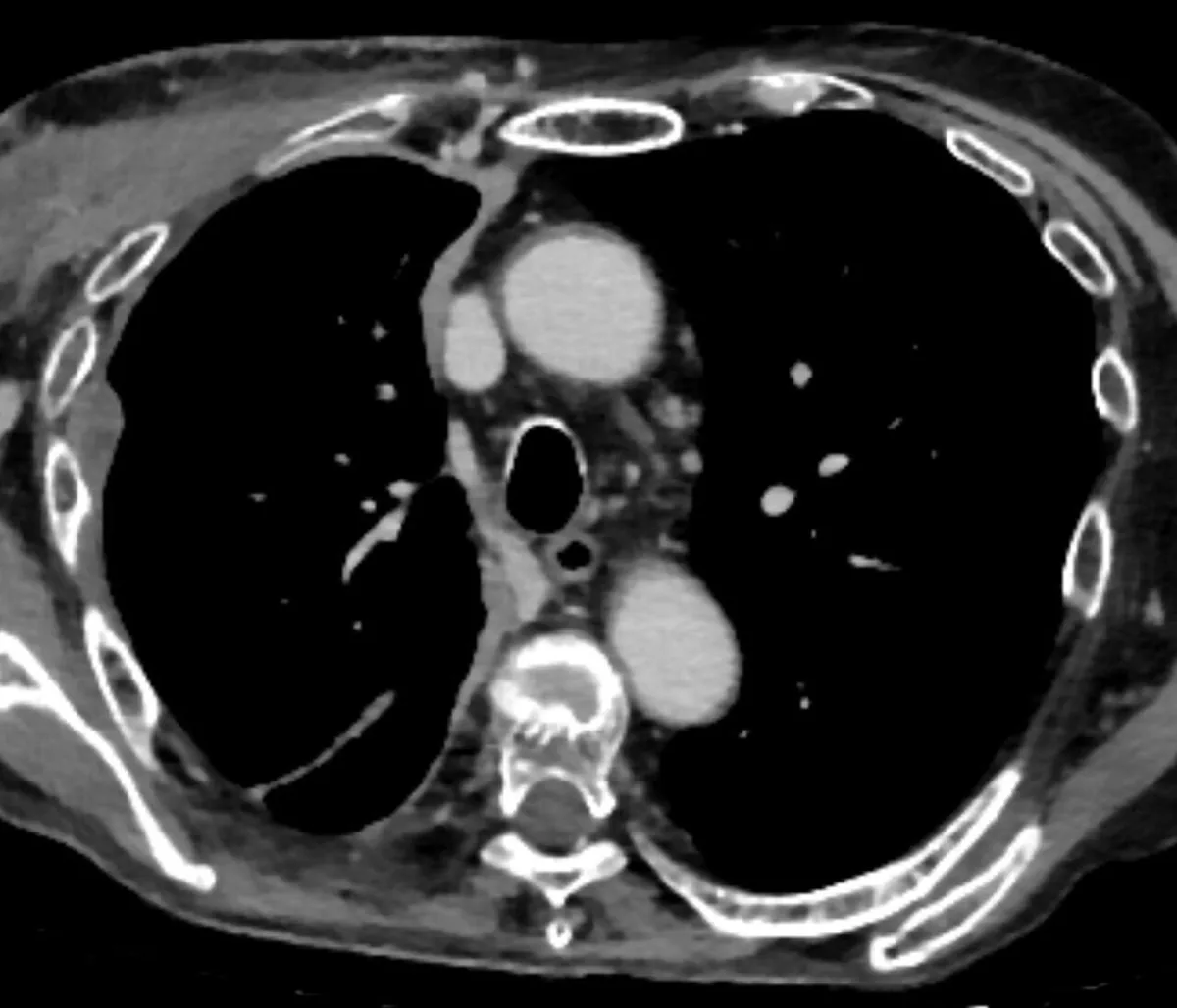 CT
CT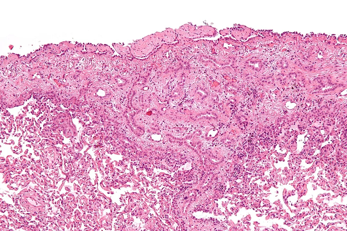 Histology
Definition
Asbestos is a group of naturally occurring silicate fibers. Asbestosis is an interstitial pulmonary fibrosis caused by asbestos. Malignant pleural mesothelioma is a rare malignant tumor of the serous membranes (mesothelium) that is caused by asbestos in the vast majority of cases. Asbestos also causes benign pleural changes.
Classification
- Asbestos fibers: serpentine (chrysotile) and amphiboles (amosite, crocidolite, anthophyllite, tremolite, actinolite); amphibole fibers generally have a higher potency for mesothelioma.
- Benign pleural diseases: pleural plaques (often calcified), diffuse pleural thickening, rounded atelectasis, benign asbestos pleural effusion; they are more common than asbestosis and develop with lower exposure.
- Asbestosis: pulmonary fibrosis.
- Malignant mesothelioma: up to 90 % in the pleura, most of the rest in the peritoneum, rarely in the pericardium or tunica vaginalis.
Occurrence & epidemiology
Direct occupational exposure is the main cause of asbestos-related disease. Particularly affected were construction trades (insulators, pipefitters, carpenters, electricians, roofers, drywall installers), shipyard workers and naval personnel, boiler and furnace builders, brake mechanics and workers in asbestos mining and processing. Today exposure occurs mainly during repair, renovation, dismantling or maintenance of older asbestos-containing materials. Mesothelioma used to occur mainly in men in industrial occupations; as incidence in men has fallen, the proportion in women has increased.
Aetiopathogenesis
- Asbestosis: alveolar macrophages attempt to phagocytose the fibers and release cytokines and growth factors that promote inflammation, oxidative damage, collagen deposition and fibrosis; the fibers can also be directly toxic. The risk depends on duration and intensity of exposure and on type, length and thickness of the fibers. The latency is usually 20–40 years.
- Pleural plaques: latency usually ≥ 20 years; a marker of substantial exposure. Benign asbestos pleural effusions occur ≥ 10 years after first exposure.
- Mesothelioma: the risk rises with exposure, but cases also occur after low exposure; the latency is 30–50 years. Besides occupational exposure, household and environmental exposure (e.g. erionite in Cappadocia) play a role. The risk is independent of smoking.
Clinical features
- Asbestosis: gradually increasing exertional breathlessness, non-productive cough and fatigue; slow progression over years, even after exposure has ended; in severe cases end-stage fibrosis and cor pulmonale.
- Pleural changes: usually asymptomatic; diffuse pleural thickening can cause symptomatic restriction.
- Mesothelioma: breathlessness and non-pleuritic chest pain; pleural effusions, usually unilateral and hemorrhagic, in up to 90 %. Invasion of the chest wall and adjacent structures causes severe pain, hoarseness, dysphagia, brachial plexopathy or ascites; general symptoms are rare at first presentation.
Histology
In mesothelioma two main subtypes are distinguished: the epithelioid type (about 60 % or more, the most common subtype) and the sarcomatoid type. The diagnosis is based on histology and immunohistochemistry; distinction from other tumors can be difficult. In asbestosis, asbestos fibers and asbestos bodies in lavage fluid or lung tissue can support the diagnosis but are not required.
Diagnosis
- Occupational and exposure history: onset, duration, type and intensity of exposure.
- Asbestosis on chest X-ray: bilateral linear reticular opacities, mostly in the peripheral lower zones, with or without pleural changes; graded according to the ILO classification of pneumoconioses. Hilar or mediastinal lymphadenopathy and a nodular pattern are atypical.
- HRCT: more sensitive for pleural and parenchymal changes; usually a UIP pattern, honeycombing in the middle and lower zones indicates advanced disease.
- Pleura: bilateral pleural plaques and/or diaphragmatic calcification are virtually pathognomonic of previous asbestos exposure; plaques involve the parietal pleura and diaphragm, sparing the apices and costophrenic angles. Rounded atelectasis appears as a curvilinear, scar-like mass, often in the lower zones.
- Lung function: restriction, reduced DLCO and/or mild obstruction.
- Mesothelioma: nodular pleural thickening, pleural effusion and volume loss of the hemithorax on X-ray; CT, MRI and PET-CT for extent. Pleural biopsy is usually required because effusion cytology is usually not diagnostic.
Keep learning in the app
Further reading (open access)
Cross-references
Note: Learning content for medical education – not a treatment recommendation and no substitute for diagnosis or treatment decisions in individual cases. Treatment and management are deliberately not covered on this page.