Vasa praevia
Exam relevance: in 4 of 197 board exam reports · rank 111
- Specialty
- Obstetrics · Pregnancy disorders
- Images
- Endoscopy & gross 1 · Clinical 1
- Exam relevance
- 4 of 197 reports · rank 111
- In the app
- 1 flashcards · GynFuchs
- Last updated
- 10/2026 · Dr. Pascal Bafteh
Contents
Images (2)
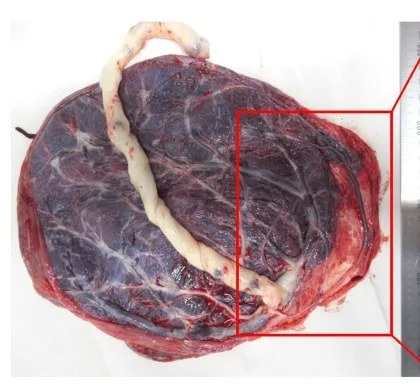 Endoscopy & gross
Definition
- Vasa praevia means fetal vessels running unprotected over or close to the internal os.
- The usual cause is a velamentous cord insertion or a succenturiate lobe with connecting vessels.
- Risk factors are a low-lying placenta, multiple pregnancy, assisted reproduction and accessory lobes.
Classification
- Type I: the unprotected vessels arise from a velamentous cord insertion.
- Type II: the vessels connect the main placenta with an accessory lobe (succenturiate or bilobed placenta) and cross the internal os.
Occurrence & epidemiology
Epidemiology
- Vasa praevia is rare.
- It is considerably more common after assisted reproduction and in multiple pregnancies.
Clinical features
- Undiagnosed vasa praevia is often asymptomatic until the membranes rupture.
- If the unprotected vessels tear at membrane rupture, painless vaginal bleeding from the fetal circulation follows, with rapid fetal bradycardia because the fetal blood volume is small.
- Compression of the vessels by the presenting part can also cause fetal heart rate changes.
Diagnosis
- It is made transvaginally with colour Doppler, often incidentally while looking at the cervix.
- The differential includes a cord loop in the lower segment, which moves when she changes position.
Imaging findings
- Transvaginal colour Doppler ultrasound shows vessels running over or immediately next to the internal os, with a fetal arterial or venous flow pattern on pulsed Doppler.
Keep learning in the app
Further reading (selection)
Cross-references
Note: Learning content from the GynFuchs app (flashcards, exam questions, image cases) for medical education – not a treatment recommendation and no substitute for diagnosis or treatment decisions in individual cases. Treatment and management are deliberately not covered on this page.