Acute fatty liver of pregnancy
- Specialty
- Obstetrics · Pregnancy disorders
- Images
- Histology & cytology 1
- Last updated
- 10/2026 · Dr. Pascal Bafteh
Contents
Images (1)
Definition
- Acute fatty liver of pregnancy is a rare, life-threatening complication of late pregnancy with acute liver dysfunction due to microvesicular fatty infiltration of the liver parenchyma, which can lead to coagulopathy, electrolyte imbalance and multi-organ failure.
Occurrence & epidemiology
Epidemiology
- The incidence is about 1 in 7,000 to 1 in 20,000 pregnancies; it recurs in a later pregnancy in about 20%.
Aetiopathogenesis
Aetiology and risk factors
- The cause is mitochondrial dysfunction and defects of fetal and placental fatty acid oxidation, especially fetal LCHAD deficiency; toxic 3-hydroxy fatty acids reach the maternal circulation and cause microvesicular fatty liver.
- Primiparous women, pregnancies with a male fetus, multiple pregnancies, women with previous acute fatty liver of pregnancy and low BMI are more often affected; pre-eclampsia coexists in more than 50%.
Clinical features
Clinical features and complications
- It occurs in the third trimester, rarely late in the second trimester or shortly after birth; initially there is non-specific epigastric pain, nausea and vomiting with raised transaminases.
- Early complications are hypoglycaemia, acute pancreatitis, infection and acute renal failure; signs of acute liver failure such as encephalopathy and bleeding from coagulopathy usually follow 1–2 weeks after onset, together with disseminated intravascular coagulation and postpartum haemorrhage.
Histology
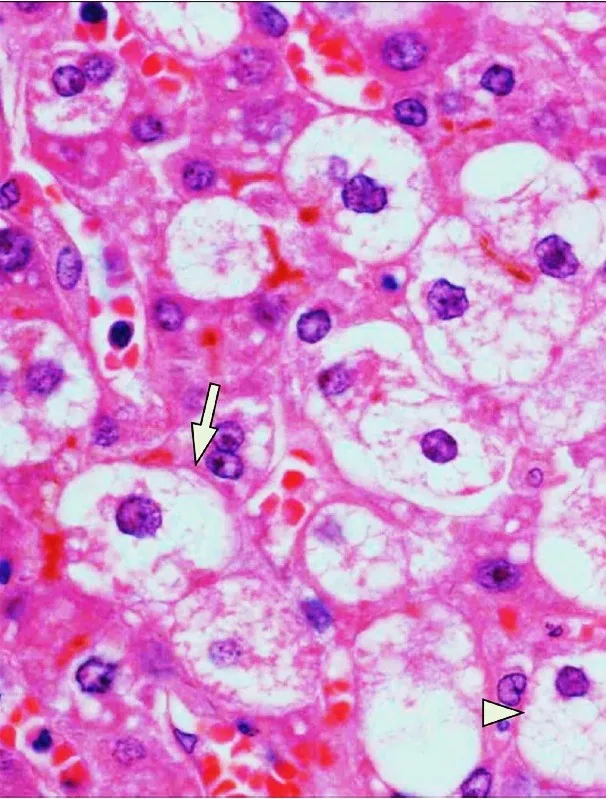
- Liver biopsy typically shows microvesicular fatty infiltration of hepatocytes; it is not required for the diagnosis and is rarely performed in pregnancy.
Diagnosis
- Laboratory findings include moderately raised transaminases that do not correlate with severity; leucocytosis and raised urea and creatinine are common, followed by hypoglycaemia and raised ammonia; platelets are usually normal unless DIC is present.
- The Swansea criteria standardise the diagnosis: at least 6 of 14 points from clinical and laboratory findings such as nausea or vomiting, abdominal pain, encephalopathy, raised bilirubin, leucocytosis, raised transaminases and creatinine, prolonged PT/aPTT and steatosis on ultrasound support the diagnosis.
- Imaging contributes little to the diagnosis and mainly serves to exclude other liver processes such as haematoma or rupture.
Keep learning in the app
Further reading (selection)
Cross-references
Note: Learning content from the GynFuchs app (flashcards, exam questions, image cases) for medical education – not a treatment recommendation and no substitute for diagnosis or treatment decisions in individual cases. Treatment and management are deliberately not covered on this page.