Placental abruption
Exam relevance: in 16 of 197 board exam reports · rank 46
- Synonyms
- abruptio placentae
- Specialty
- Obstetrics · Pregnancy disorders
- Images
- Ultrasound 2 · Clinical 1
- Exam relevance
- 16 of 197 reports · rank 46
- In the app
- 2 flashcards · GynFuchs
- Last updated
- 10/2026 · Dr. Pascal Bafteh
Contents
Images (3)
 Ultrasound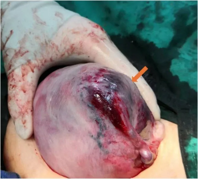
 Ultrasound
Aetiopathogenesis
Risk factors and key findings
- Cardinal finding: painful bleeding with a hard, tender uterus and a pathological CTG.
- The dangerous variant is concealed bleeding with no visible loss — the shock does not match what you can see.
- Risk factors: hypertension and pre-eclampsia, trauma, smoking and cocaine, ruptured membranes, polyhydramnios, previous abruption.
- Ultrasound signs: a hypoechoic mass between placenta and uterine wall, an undulating or sunken placental contour; importantly a normal scan does not exclude abruption.
Clinical features
Presentation
- The cardinal features are continuous pain, a board-like tender uterus and usually dark vaginal bleeding.
- In some cases the bleeding is concealed, because the haematoma collects behind the placenta — the loss is then underestimated.
- The CTG typically shows reduced variability, variable or late decelerations, progressing to bradycardia.
Complications
- The feared ones are intrauterine death and disseminated intravascular coagulation.
- The Couvelaire uterus denotes bleeding into the uterine muscle with blue-violet mottling (uteroplacental apoplexy).
- Further consequences are acute kidney injury, shock and postpartum haemorrhage.
Diagnosis
Ultrasound
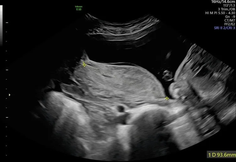
- You may see a hypoechoic or heterogeneous mass between placenta and uterine wall, and a wavy, sunken placental contour.
- What matters: a normal scan does not exclude abruption.
- Fresh haematomas can look isoechoic to the placenta and so be missed.
- The diagnosis is therefore primarily clinical.
Typical imaging findings
- Ultrasound: Between the placenta and the uterine wall a hypoechoic to mixed-echogenic area lifts the placenta marginally off the wall. The placenta appears thickened at this point; the measurement records the extent of the haematoma.
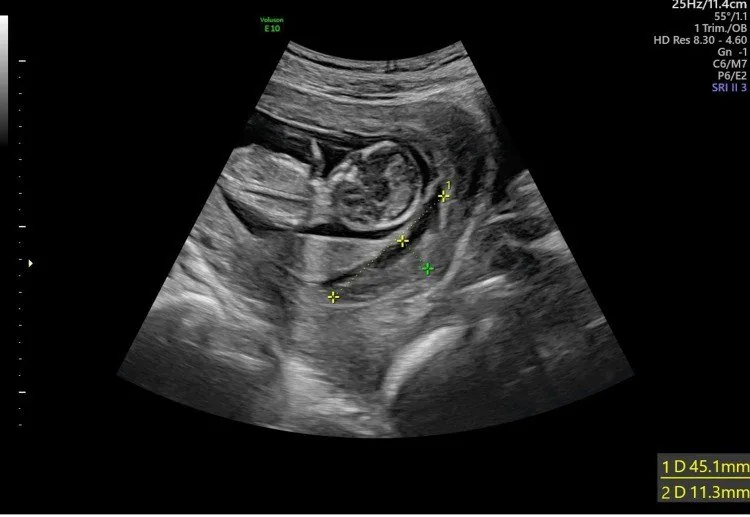
- Ultrasound: Beside the gestational sac lies a crescent-shaped hypoechoic zone lifting the chorion off the uterine wall. The sac itself is normally configured; calipers record the extent of the bleed.
Keep learning in the app
Further reading (selection)
Cross-references
Note: Learning content from the GynFuchs app (flashcards, exam questions, image cases) for medical education – not a treatment recommendation and no substitute for diagnosis or treatment decisions in individual cases. Treatment and management are deliberately not covered on this page.